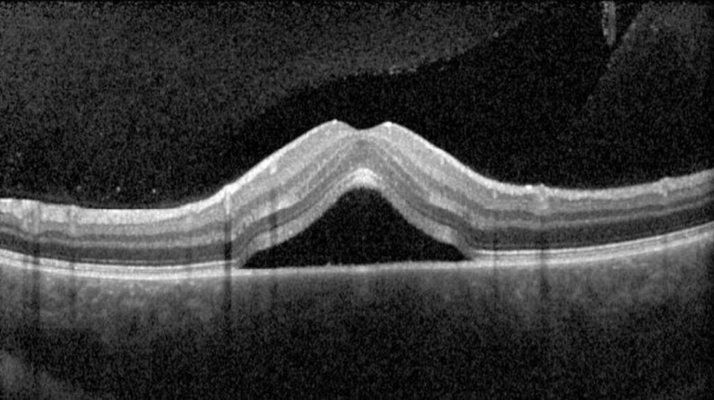
Subretinal fluid on OCT is a dark, hyporeflective space between the neurosensory retina and the retinal pigment epithelium.
It is also one of the commonest findings in retinal practice, and it appears in many diseases that need very different treatments.
Reading the context around the fluid is also what turns the finding into a diagnosis.
What Is Subretinal Fluid on OCT?
Subretinal fluid accumulates when fluid enters the potential space faster than the retinal pigment epithelium can pump it out.
In addition, the sources include leakage from choroidal vessels through a defective pigment epithelium, exudation from abnormal vessels, and passage of liquefied vitreous through a retinal break.
The fluid separates the photoreceptors from the pigment epithelium, which interrupts the visual cycle and, if prolonged, leads to photoreceptor damage.
On OCT, the detached retina is elevated over an optically empty space, and the outer segments may also appear elongated.
Subretinal Fluid on OCT: Patterns That Point to a Cause
- Central serous chorioretinopathy: a smooth serous detachment, often with a small pigment epithelial detachment and thick choroid (see central serous chorioretinopathy)
- Neovascular AMD: fluid with drusen, a pigment epithelial detachment, hyperreflective material, and a membrane (see macular neovascularization types)
- Polypoidal choroidal vasculopathy: fluid with notched or peaked pigment epithelial detachments (see polypoidal choroidal vasculopathy)
- Rhegmatogenous detachment: a mobile, corrugated retina with a break and no pigment epithelial changes (see rhegmatogenous retinal detachment)
- Inflammatory disease, such as Vogt-Koyanagi-Harada disease and posterior scleritis, with multiple pockets and thickened choroid (see Vogt-Koyanagi-Harada disease)
- Optic disc pit maculopathy with fluid that connects to the disc (see optic pit maculopathy)
- Hypertensive and preeclamptic choroidopathy (see preeclampsia retinal changes)
- Choroidal tumors and metastases, where fluid overlies a mass (see choroidal metastases)
Fundus Explorer Pro
Photograph the retinal findings described here with the phone already in your pocket — 22 D optics and built-in illumination in one handheld unit.
From Choroida — the team behind this siteFeatures of the Fluid Itself
- Clear, hypo-reflective fluid suggests serous fluid, as in CSC
- Hyperreflective dots or material within the fluid suggest fibrin, lipid, or hemorrhage, as in neovascular disease
- Shaggy photoreceptor outer segments appear in chronic detachment, with changes in the ellipsoid zone
- Fluid with intraretinal cysts suggests a chronic or exudative process
- Septated or loculated fluid suggests inflammation or a tumor
Look also at the pigment epithelium for detachments, tears, and irregularities, and at the choroid for thickness.
Complementary Tests
- Fluorescein angiography to see the source of leakage (see fluorescein angiography patterns)
- Indocyanine green angiography for choroidal vessels and polyps (see indocyanine green angiography)
- OCT angiography for neovascular membranes (see OCT angiography)
- Fundus autofluorescence for chronicity and pigment epithelial changes (see fundus autofluorescence)
- B-scan and examination of the periphery for a break or tumor
When Fluid Can Be Tolerated
In neovascular AMD, some patients with a small amount of stable subretinal fluid do as well as those with a dry macula.
In addition, the FLUID study randomized patients on a treat-and-extend regimen to a strict approach that tolerated no fluid or a more relaxed approach that tolerated some subretinal fluid, and found similar vision with fewer injections in the tolerant group (see the references).
This applies to subretinal fluid in neovascular AMD, and not to intraretinal fluid, which usually needs treatment.
In CSC, observation is the first step for acute disease, as fluid often resolves by itself.
Subretinal Fluid on OCT: Management by Cause
Treatment is directed to the cause.
- Neovascular disease: anti-VEGF therapy (see anti-VEGF therapy)
- Chronic CSC: photodynamic therapy, micropulse laser, or other options (see micropulse laser)
- Retinal detachment: surgery (see scleral buckle versus vitrectomy)
- Inflammation: steroids and immunosuppression
- Tumor: treatment of the tumor
Also, explain to the patient the cause of the fluid and what is planned.
Serial Imaging
The behavior of fluid over time gives diagnostic information: acute central serous fluid often resolves within months, whereas fluid that persists or recurs may signal chronic disease or neovascularization.
Also, measure the height of the fluid and the pigment epithelial detachment at each visit, and note any new hyperreflective material.
In treated neovascular AMD, track whether the fluid is stable, increasing, or fluctuating, since this determines the interval and the strategy.
Patient Information
Explain to the patient what the dark space on the scan means, and show them the images if possible.
In addition, describe the plan: observation, injections, laser, or surgery, and explain the likely time course.
Also, ask them to report any new distortion or loss of vision.
In every case, record whether the fluid involves the fovea, since this affects acuity and urgency, and compare with the fellow eye for a pattern that suggests a bilateral disease such as pachychoroid spectrum disease.
For the latest studies, see the PubMed results for subretinal fluid on OCT.


Document what you see
Two smartphone imaging tools built for everyday clinic use — one for the slit lamp, one for the fundus.
From Choroida — the team behind this siteReferences
- Guymer RH, Markey CM, McAllister IL, et al. Tolerating subretinal fluid in neovascular age-related macular degeneration treated with ranibizumab using a treat-and-extend regimen: FLUID study 24-month results. Ophthalmology. 2019;126:723-734.
- Spaide RF, Jaffe GJ, Sarraf D, et al. Consensus nomenclature for reporting neovascular age-related macular degeneration data: consensus on neovascular AMD nomenclature study group. Ophthalmology. 2020;127:616-636.
- Ryan SJ, Schachat AP, Wilkinson CP, et al, eds. Retina. 6th ed. Elsevier; 2018.
- American Academy of Ophthalmology. Basic and Clinical Science Course, Section 12: Retina and Vitreous. AAO; current edition.

Writes and reviews the clinical articles on Educate. Every post is checked against peer-reviewed sources before it goes live.