
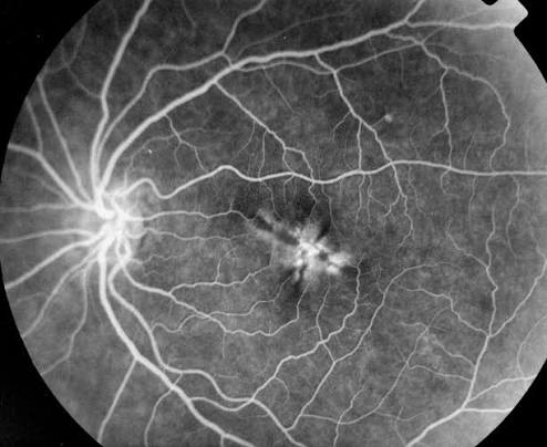
Fluorescein angiography (FA) records the passage of sodium fluorescein through the retinal and choroidal circulations using blue excitation light and a yellow-green barrier filter.
It remains the standard test for many retinal vascular and inflammatory diseases, even as OCT and OCT angiography take over parts of its role.
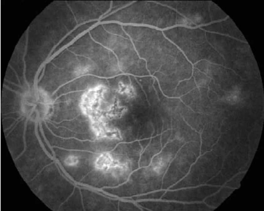
Reading an angiogram well means knowing the normal phases and then classifying abnormalities as hyperfluorescent or hypofluorescent.
This article covers the principles, the patterns, and the practical safety points.
Principles of Fluorescein Angiography
Sodium fluorescein is a small molecule that absorbs blue light at about 465 to 490 nm and emits yellow-green light at about 520 to 530 nm.
After intravenous injection, most of the dye binds to serum albumin and stays within intact vessels, and a small unbound fraction is free to leak.
The retinal capillaries have tight endothelial junctions, and the RPE forms the outer blood-retinal barrier, so normal retinal vessels do not leak.
The choriocapillaris is fenestrated, and dye leaks from it into the choroidal stroma, but the RPE blocks further passage into the retina.
Excitation and barrier filters in the fundus camera ensure that only fluorescence from the dye is photographed.
A typical dose is 500 mg of fluorescein sodium, given as 5 mL of a 10% solution or 3 mL of a 25% solution, injected into an antecubital vein.
Phases of a Normal Angiogram
- Choroidal phase: choroidal filling begins about 10 to 15 seconds after injection, with a patchy, blotchy pattern, and a cilioretinal artery, if present, fills at the same time
- Arterial phase: dye enters the retinal arteries within about a second of choroidal filling
- Arteriovenous or capillary phase: dye fills the capillaries and the foveal avascular zone becomes visible as a dark area
- Venous phase: laminar flow along the vein walls, followed by complete venous filling
- Late phase: dye recirculates and fades, and at 10 minutes or more only staining of the disc, sclera, and some lesions persists
A delay in choroidal or retinal filling is abnormal and suggests carotid disease, ocular ischemia, or arterial occlusion.
Fundus Explorer Pro
Photograph the retinal findings described here with the phone already in your pocket — 22 D optics and built-in illumination in one handheld unit.
From Choroida — the team behind this siteHypofluorescence
Hypofluorescence means less fluorescence than expected.
Blocked Fluorescence
Material in front of the vessels or RPE blocks the view.
- Retinal or preretinal hemorrhage
- Pigment, such as a nevus, CHRPE, or RPE hyperplasia
- Lipid exudates
- Vitreous opacities and media haze
Blocked fluorescence stays dark throughout the study, and the shape corresponds to the lesion on color photography.
Vascular Filling Defects
Vessels do not fill because of occlusion or nonperfusion.
- Retinal arterial or venous occlusion
- Capillary nonperfusion in diabetic retinopathy and vein occlusion
- Choroidal ischemia and choriocapillaris loss
- Optic disc hypoperfusion
Areas of capillary dropout appear dark, with abrupt margins.
Hyperfluorescence
Hyperfluorescence means more fluorescence than expected.
Window Defects
Loss of the RPE lets the choroidal fluorescence show through.
- Atrophy, as in geographic atrophy and macular scars
- Drusen, which stain early and fade in the late phase
- Old RPE tears and laser scars
A window defect appears early, reaches maximum intensity by the venous phase, and fades without increase in size.
Leakage
Dye escapes from abnormal vessels or from damaged barrier.
- Neovascularization: intense, progressive leakage, seen in retinal and disc neovascularization, and in classic choroidal neovascularization
- Microaneurysms: punctate hyperfluorescent dots that may leak in diabetic retinopathy
- Macular edema: petaloid or diffuse pooling in cystoid spaces, with late disc leakage in some eyes (see cystoid macular edema)
- Vasculitis: leakage and staining along vessel walls
- Central serous chorioretinopathy: an expanding ink-blot or smokestack leak (see central serous chorioretinopathy)
Pooling
Dye collects in a space with defined borders.
- Pigment epithelial detachment fills early and stays bright and sharply demarcated
- Serous retinal detachment fills slowly and progressively in the subretinal space
Staining
Tissue takes up dye, and the hyperfluorescence persists in the late phase without change in shape.
- Drusen, scars, and fibrous tissue
- The optic disc and sclera
Recognizing which pattern is present is often more useful than the exact diagnosis, and the pattern narrows the list of possibilities (see leaky vessels on angiography).
Practical Diagnostic Uses
- Diabetic retinopathy: microaneurysms, macular leakage, capillary dropout, and neovascularization
- Vein occlusion: delayed venous filling, nonperfusion, and leakage
- Neovascular AMD: classic and occult patterns, now replaced by anatomic types
- Inflammatory disease: vasculitis, disc leakage, and choroiditis lesions
- Tumors: hyperfluorescence patterns and leak
- Central serous chorioretinopathy: site of leakage, to guide treatment
Widefield angiography extends these observations to the periphery.
OCT angiography complements FA by showing vascular detail without dye (see OCT angiography).
Safety and Adverse Effects
Fluorescein is generally safe, and most reactions are mild.
- Nausea is the most common reaction, sometimes with vomiting, and it usually settles quickly
- Skin and urine turn yellow for up to 24 to 36 hours
- Urticaria and itching occur in some patients
- Extravasation causes local pain and may need cold compresses
- Vasovagal reactions and syncope occur
- Severe reactions such as bronchospasm, laryngeal edema, and anaphylaxis are rare, and the clinic must be equipped to treat them
Prospective surveys have found that mild reactions are common and severe reactions are rare.
A history of a prior severe reaction is a contraindication.
In pregnancy, fluorescein is used only when clearly necessary, and OCT and OCT angiography are alternatives.
Patients with renal disease can generally receive the standard dose, although they may have prolonged yellow discoloration.
Patients on beta-blockers need attention, since anaphylaxis may be harder to treat.


Document what you see
Two smartphone imaging tools built for everyday clinic use — one for the slit lamp, one for the fundus.
From Choroida — the team behind this siteReferences
- Novotny HR, Alvis DL. A method of photographing fluorescence in circulating blood in the human retina. Circulation. 1961;24:82-86.
- Yannuzzi LA, Rohrer KT, Tindel LJ, et al. Fluorescein angiography complication survey. Ophthalmology. 1986;93:611-617.
- Kwiterovich KA, Maguire MG, Murphy RP, et al. Frequency of adverse systemic reactions after fluorescein angiography: results of a prospective study. Ophthalmology. 1991;98:1139-1142.
- Lopez-Saez MP, Ordoqui E, Tornero P, et al. Fluorescein-induced allergic reaction. Ann Allergy Asthma Immunol. 1998;81:428-430.
- Gass JDM. Stereoscopic Atlas of Macular Diseases: Diagnosis and Treatment. 4th ed. St Louis: Mosby; 1997.
Test yourself
A few questions straight from this article.
-
Sodium fluorescein absorbs blue light and then emits light of what colour?
Fluorescein absorbs blue light at about 465 to 490 nm and emits yellow-green light at about 520 to 530 nm; the camera's excitation and barrier filters ensure only this fluorescence is photographed. -
Why do normal retinal vessels not leak fluorescein?
Retinal capillaries have tight endothelial junctions and the RPE forms the outer blood-retinal barrier, so normal retinal vessels do not leak. Most dye is albumin-bound, but a small unbound fraction is free to leak. -
When does choroidal filling begin on a normal fluorescein angiogram?
Choroidal filling begins about 10 to 15 seconds after injection with a patchy, blotchy pattern, and the retinal arteries fill within about a second afterwards. -
How does a window defect behave across a fluorescein angiogram?
Loss of RPE lets choroidal fluorescence show through, so a window defect appears early, reaches maximum intensity by the venous phase, and fades without increasing in size. -
Which angiographic pattern shows hyperfluorescence that persists late without changing shape?
Staining is uptake of dye by tissue such as scars, fibrous tissue, the optic disc and sclera, and the hyperfluorescence persists into the late phase without change in shape. -
Which finding produces blocked fluorescence on an angiogram?
Material in front of the vessels or RPE blocks the view, including retinal and preretinal hemorrhage, pigment, lipid exudates and media opacities. Blocked fluorescence stays dark throughout, matching the lesion on colour photography. -
How does a pigment epithelial detachment appear on fluorescein angiography?
Pooling means dye collecting in a space with defined borders. A pigment epithelial detachment fills early and stays bright and sharply demarcated, whereas a serous retinal detachment fills slowly and progressively. -
Which leakage pattern is characteristic of central serous chorioretinopathy?
Central serous chorioretinopathy produces an expanding ink-blot or smokestack leak, and angiography is used to locate the leak point to guide treatment. -
What is the most common adverse reaction to intravenous fluorescein?
Nausea is the most common reaction and usually settles quickly. Urticaria and vasovagal reactions also occur, while bronchospasm, laryngeal edema and anaphylaxis are rare but need a clinic equipped to treat them. -
Which history is a contraindication to fluorescein angiography?
A history of a prior severe reaction is a contraindication. Patients with renal disease can generally receive the standard dose, and beta-blocker use simply calls for extra attention since anaphylaxis may be harder to treat.

Writes and reviews the clinical articles on Educate. Every post is checked against peer-reviewed sources before it goes live.


