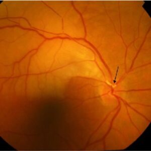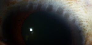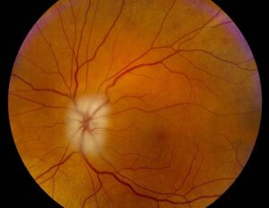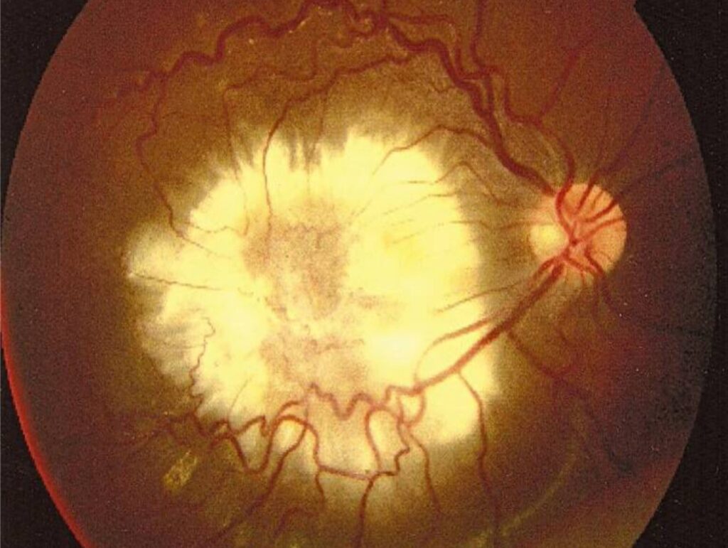
Ocular toxocariasis results from a single migrating larva becoming trapped within the eye, and this unilateral, single-organism nature explains why the disease looks so different from a typical infection: rather than a diffuse inflammatory process, it produces a localized granulomatous reaction wherever that one larva happened to come to rest, giving rise to several distinct, location-dependent clinical presentations that all fall under the same underlying diagnosis.
The Parasite and How the Eye Becomes Involved
Toxocara canis, and less commonly Toxocara cati, are roundworm parasites of dogs and cats, and human infection occurs through accidental ingestion of infective eggs, most often in young children with a history of exposure to contaminated soil or close contact with puppies, particularly given typical hand-to-mouth behavior at this age.
In ocular toxocariasis, a single larva migrates to and becomes trapped within the eye, where it eventually dies, provoking a localized granulomatous inflammatory response around the dead organism, in contrast to visceral larva migrans, in which larvae are more widely disseminated through other organs.
Clinical Presentations
Posterior Pole (Localized) Granuloma
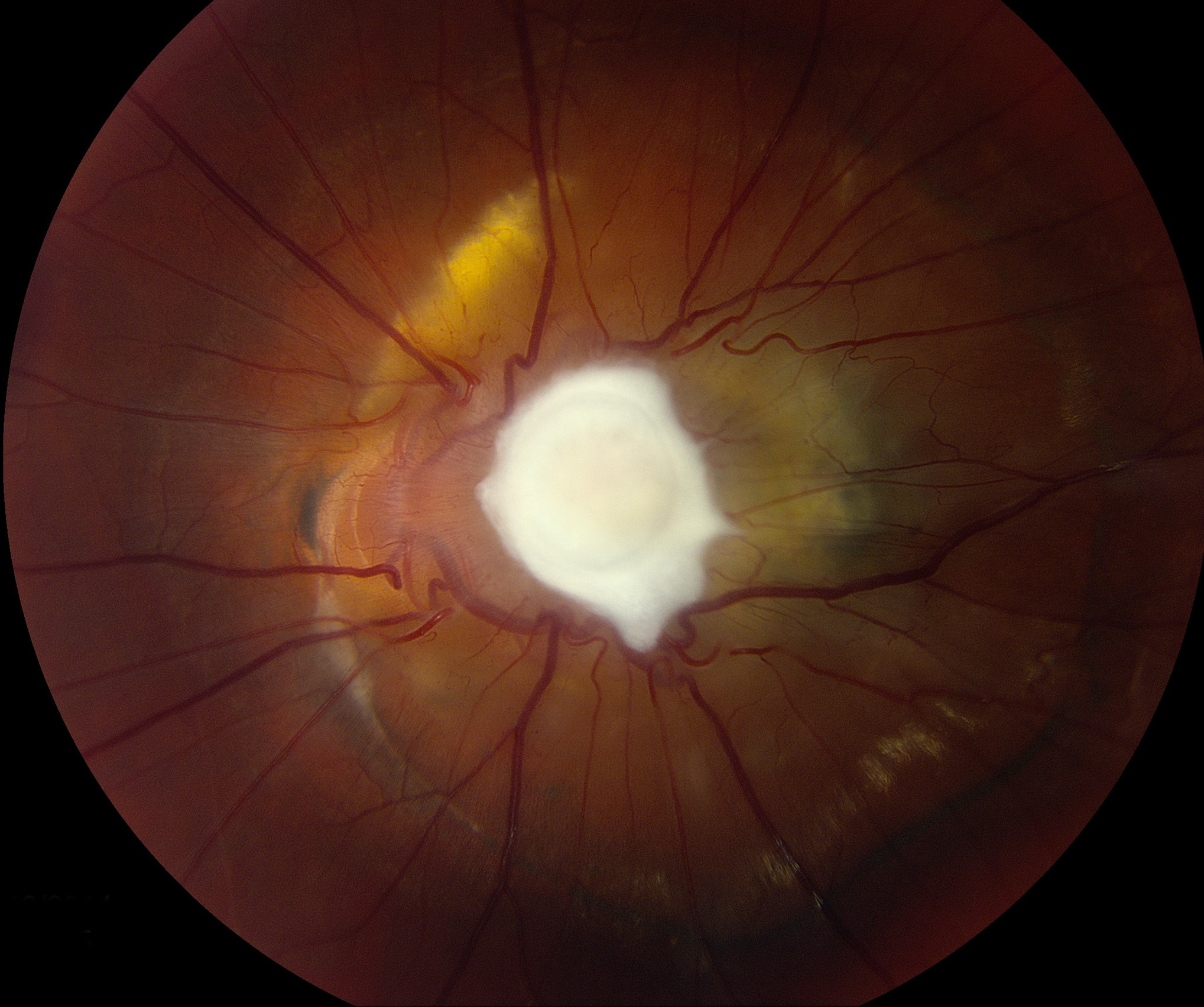
A discrete, elevated, white to yellow granulomatous mass, typically located at or near the posterior pole, often associated with some degree of vitreous traction and, in some cases, a tractional band extending toward the optic disc or periphery.
Peripheral Granuloma
A similar granulomatous mass located in the peripheral retina, sometimes associated with a tractional retinal fold extending toward the macula or optic disc, which can itself cause visually significant macular distortion even though the primary granuloma is peripherally located.
Chronic Endophthalmitis
A more diffuse pattern of chronic intraocular inflammation, with vitritis and, over time, potential complications including cataract, band keratopathy, and hypotony, generally representing a more severe or advanced presentation than the localized granuloma forms.
Fundus Explorer Pro
Photograph the retinal findings described here with the phone already in your pocket — 22 D optics and built-in illumination in one handheld unit.
From Choroida — the team behind this siteWhy It’s Confused With Retinoblastoma
Like Coats disease, ocular toxocariasis typically presents in a similar age group to retinoblastoma and can cause leukocoria or strabismus, particularly with a posterior pole granuloma or significant associated vitritis and traction, making this differential distinction, alongside Coats disease, one of the essential considerations in any child presenting with a white pupillary reflex (see Coats-like retinal telangiectasia for the related distinguishing considerations).
Diagnostic Evaluation
- Careful dilated fundus examination characterizing the location, appearance, and any associated traction related to the granuloma
- A history of exposure risk factors, including contact with puppies or contaminated soil, though a definitive exposure history is not always obtainable or necessary for diagnosis
- Serologic testing for Toxocara antibodies, using enzyme-linked immunosorbent assay, supports the diagnosis, though interpretation requires clinical correlation given the possibility of prior asymptomatic exposure in the general population
- Ultrasound and, when the diagnosis remains uncertain, other imaging help characterize the lesion and exclude a solid tumor mass consistent with retinoblastoma, particularly important given the shared age group and potential for overlapping presentation
- Systemic evaluation for visceral larva migrans is generally not necessary in isolated ocular toxocariasis, since ocular disease typically represents an isolated single-larva event rather than evidence of broader systemic dissemination
Management
Anti-Inflammatory Treatment
Corticosteroids, topical for anterior segment inflammation and periocular or systemic for more significant posterior segment involvement, are central to managing the inflammatory response, since the visual damage in ocular toxocariasis results primarily from the host inflammatory reaction to the dead larva rather than from the organism itself, which is already dead by the time most patients present.
Antiparasitic Therapy
The role of antiparasitic medication is more limited than in some other parasitic ocular infections, given that the causative larva has typically already died by the time of diagnosis, though antiparasitic treatment may be considered in select cases.
Surgical Management
Vitrectomy can be considered for eyes with significant vitreous traction, tractional retinal detachment, or dense vitreous opacity significantly limiting vision, aiming to relieve mechanical complications of the chronic granulomatous and tractional process.
Prognosis
Visual outcomes in ocular toxocariasis depend heavily on the location of the granuloma, with posterior pole and macula-involving lesions or significant associated traction carrying a more guarded visual prognosis than peripheral lesions without central macular involvement.
Prompt, correct diagnosis, distinguishing this condition from retinoblastoma, is essential both to direct the correct treatment for toxocariasis itself and, just as importantly, to avoid either missing a true retinoblastoma or subjecting a child with toxocariasis to an unnecessary and invasive workup intended for a malignant tumor.


Document what you see
Two smartphone imaging tools built for everyday clinic use — one for the slit lamp, one for the fundus.
From Choroida — the team behind this siteReferences
- Shields JA. Ocular toxocariasis. A review. Surv Ophthalmol. 1984;28:361-381.
- Despreaux R, Fardeau C, Touhami S, et al. Ocular toxocariasis: clinical features and long-term visual outcomes in adult patients. Am J Ophthalmol. 2016;166:162-168.
- Stewart JM, Cubillan LD, Cunningham ET Jr. Prevalence, clinical features, and causes of vision loss among patients with ocular toxocariasis. Retina. 2005;25:1005-1013.
- Shields CL, Shields JA. Ocular toxocariasis simulating retinoblastoma. Br J Ophthalmol. 1990;74:335-338.
Test yourself
A few questions straight from this article.
-
Which parasite causes ocular toxocariasis, and in which animal does it normally live?
Toxocara canis, and less commonly Toxocara cati, are roundworm parasites of dogs and cats that can infect humans accidentally. -
How do humans acquire Toxocara infection?
Infection follows accidental ingestion of infective eggs, most often in young children exposed to contaminated soil or puppies, given typical hand-to-mouth behaviour at that age. -
Why does ocular toxocariasis produce a localized lesion rather than diffuse infection?
One larva migrates into the eye, becomes trapped and dies there, and the surrounding granulomatous reaction forms wherever that single organism came to rest, unlike the widespread dissemination of visceral larva migrans. -
What makes a peripheral Toxocara granuloma visually significant?
A peripheral granuloma can drag a tractional retinal fold towards the macula or optic disc, causing visually significant macular distortion even though the lesion itself is peripheral. -
Which complications arise in the chronic endophthalmitis form of ocular toxocariasis?
The chronic endophthalmitis pattern brings diffuse intraocular inflammation with vitritis and, over time, cataract, band keratopathy, and hypotony; it is a more severe presentation than the localized granulomas. -
Why is ocular toxocariasis confused with retinoblastoma?
Toxocariasis presents in a similar age group to retinoblastoma and can cause leukocoria or strabismus, especially with a posterior pole granuloma or marked vitritis and traction. -
Which laboratory test supports a diagnosis of ocular toxocariasis?
Serologic testing by ELISA supports the diagnosis, but results need clinical correlation because asymptomatic prior exposure is common in the general population. -
Why are corticosteroids central to the treatment of ocular toxocariasis?
The larva is usually already dead by presentation, so damage comes from the host inflammatory reaction; steroids are given topically for anterior inflammation and periocularly or systemically for posterior involvement. -
In which situation is vitrectomy considered in ocular toxocariasis?
Surgery is aimed at relieving the mechanical consequences of the chronic granulomatous and tractional process when traction, tractional detachment, or dense vitreous opacity limits vision. -
Which Toxocara granuloma location carries the more guarded visual prognosis?
Posterior pole and macula-involving granulomas, or those with significant associated traction, carry a more guarded prognosis than peripheral lesions that spare the central macula.

Writes and reviews the clinical articles on Educate. Every post is checked against peer-reviewed sources before it goes live.