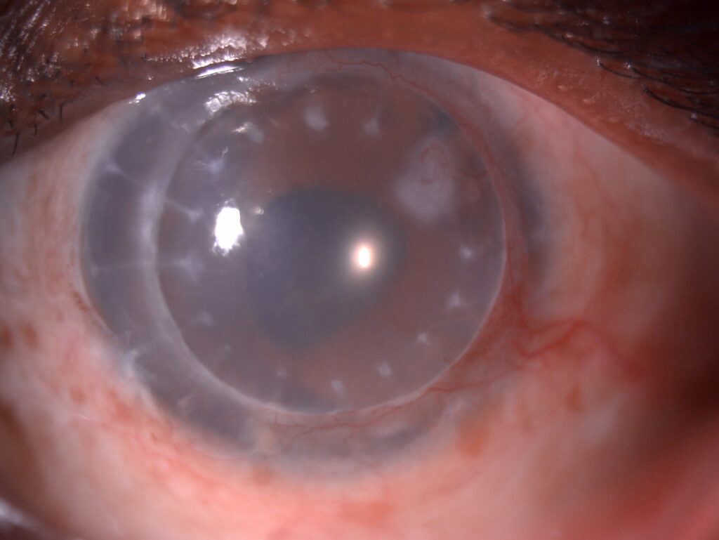
Post-keratoplasty astigmatism is the irregular or regular corneal astigmatism that follows corneal transplantation.
It is the most common reason for reduced vision in a graft that is otherwise clear.
Averages of several diopters are common after penetrating keratoplasty, and the cylinder can be high and irregular.
A structured approach, which begins with the sutures and continues through topography-guided options, usually brings most patients to useful vision.
What Causes Post-Keratoplasty Astigmatism?
The graft-host junction is a wound, and the cornea responds to it in a way that changes its curvature.
Factors include the tension of the sutures, the uneven distribution of tension around the circumference, and the difference in thickness and shape between donor and host.
Oblique or decentered trephination and a tilted graft add to the problem, as do wound healing differences between quadrants.
After endothelial keratoplasty, the astigmatism is much lower, since the surgeon does not incise the surface.
This is one reason for the shift toward endothelial procedures (see DSEK and DMEK).
Deep anterior lamellar keratoplasty has astigmatism similar to or slightly lower than penetrating keratoplasty (see DALK).
How It Presents
- Blurred and distorted vision with a clear graft and a good endothelium
- Poor correction with spectacles, because the astigmatism is irregular
- Monocular diplopia or ghost images
- Contact lens intolerance after a period of good wear
- A topography map showing a steep axis, often an asymmetric bow tie
Measure the manifest refraction, keratometry, and topography at each visit, and look for the steep axis that identifies the tight sutures.
All-fit Slit-Lamp Adapter
Record and share exactly what you see at the slit lamp. One adapter fits any slit lamp or surgical microscope — and any smartphone.
From Choroida — the team behind this sitePost-Keratoplasty Astigmatism: Management in the Early Period
Most surgeons manage astigmatism with sutures in the first months.
- Selective suture removal: surgeons remove interrupted sutures along the steep meridian to flatten that axis
- Adjustment of a running suture: surgeons can change tension in the operating room or at the slit lamp in the early weeks
- Timing: keratometry and topography guide suture removal, which usually begins a few months after surgery, with all sutures out by one year in most patients
After each removal, repeat the measurement after several weeks, because the effect develops gradually.
Patients need counselling about the risk of wound dehiscence and of infection or rejection after suture removal.
They should report pain or redness at once (see corneal graft rejection).
Post-Keratoplasty Astigmatism: Management After the Sutures Are Out
When the refraction stays stable for several months and the sutures are out, additional options are available.
- Spectacles and contact lenses: rigid gas-permeable and scleral lenses correct irregular astigmatism well (see scleral contact lens fitting)
- Arcuate and relaxing incisions: placed in the graft or at the graft-host junction, often with a femtosecond laser
- Compression sutures or wedge resection: for high astigmatism that does not respond to other measures
- Excimer laser surface ablation or LASIK: after stability, with caution over weakening the graft
- Toric intraocular lens: an option when cataract surgery is needed
Femtosecond-assisted arcuate incisions have a predictable effect and can be repeated, and they reduce the cylinder in many patients.
Excimer laser procedures deserve consideration only when the cornea is stable, the graft is healthy, and you have counselled the patient about the risk of ectasia and rejection.
Practical Pearls
Wait until the refraction is stable before deciding on laser or incisions, which may take a year or more.
Check the endothelial cell count and the pachymetry before any surgery that disturbs the graft.
Ask the patient about the visual demands, because some need near-perfect correction and others are satisfied with a contact lens.
Remember that sutures may loosen and cause infection or vascularization, so remove loose sutures promptly.
Measuring Astigmatism After a Graft
Standard keratometry is often unreliable in a grafted cornea, because the mires may be distorted or the graft may be steep and irregular.
Topography gives the shape of the whole graft and helps to decide which sutures are tight, which is the basis of selective suture removal.
Compare the keratometric axis with the refractive axis, and remove sutures only when the topography and refraction agree on the steep meridian.
Rigid Lenses and Scleral Lenses
Many grafted corneas are irregular, and a rigid lens neutralizes the irregular surface better than spectacles.
Scleral lenses vault the graft and avoid contact with the graft-host junction, which can be an advantage in eyes at risk of rejection or with a sensitive surface.
Fitting should be done by an experienced practitioner, and the lens should be checked regularly for tolerance and for any effect on the endothelium.
Record the date of each suture removal and the topography before and after, so that the response can be reviewed and the plan can be adjusted if the cylinder does not behave as expected.
For the latest studies, see the PubMed results for post-keratoplasty astigmatism.


Document what you see
Two smartphone imaging tools built for everyday clinic use — one for the slit lamp, one for the fundus.
From Choroida — the team behind this siteReferences
- Riddle HK Jr, Parker DS, Price FW Jr. Management of postkeratoplasty astigmatism. Curr Opin Ophthalmol. 1998;9:15-28.
- Feizi S, Zare M. Current approaches for management of postpenetrating keratoplasty astigmatism. J Ophthalmol. 2011;2011:708736.
- Mannis MJ, Holland EJ, eds. Cornea. 4th ed. Elsevier; 2017.
- American Academy of Ophthalmology. Basic and Clinical Science Course, Section 8: External Disease and Cornea. AAO; current edition.

Writes and reviews the clinical articles on Educate. Every post is checked against peer-reviewed sources before it goes live.