
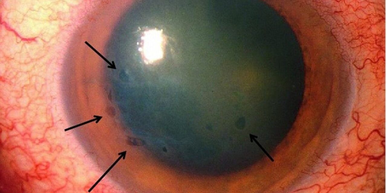
Aphakic bullous keratopathy describes corneal decompensation occurring in an eye without an intraocular lens, and while it was once a far more common complication from an era when cataract surgery routinely left eyes aphakic, it remains clinically relevant today, encountered specifically in eyes that lost or never received an intraocular lens due to surgical complications, trauma, or, less commonly now, deliberate management decisions.
Why Aphakic Eyes Are More Vulnerable to Endothelial Decompensation
In an aphakic eye, without the normal intraocular lens acting as a partial physical barrier, the vitreous can more readily move forward and make direct or near-direct contact with the corneal endothelium, particularly with eye movement or in the setting of a disrupted posterior capsule, a phenomenon sometimes called vitreous touch syndrome, which can mechanically and chemically stress and damage endothelial cells over time.
Aphakic eyes, historically resulting from intracapsular cataract extraction (a technique that removes the entire lens, including its capsule, leaving no capsular support at all) were particularly predisposed to this problem, given the complete absence of any capsular barrier between the vitreous and the corneal endothelium.
Current Clinical Contexts
While intracapsular cataract extraction is now rarely performed in modern practice, aphakia and its associated bullous keratopathy risk still occur in specific contemporary situations.
- Eyes that experienced a significant intraoperative complication during cataract surgery precluding safe intraocular lens placement at the time of the original procedure
- Eyes that have had a prior intraocular lens removed due to a complication, such as dislocation, infection, or intolerance, without immediate lens replacement
- Traumatic loss of the lens without subsequent lens implantation
- Some pediatric cataract surgery cases, where intraocular lens implantation is sometimes deferred or avoided in very young infants given the specific considerations relevant to this age group (see pediatric cataract surgery for the broader discussion of lens implantation timing considerations in this population)
All-fit Slit-Lamp Adapter
Record and share exactly what you see at the slit lamp. One adapter fits any slit lamp or surgical microscope — and any smartphone.
From Choroida — the team behind this siteClinical Features
- Progressive corneal edema and eventual bullous change (painful epithelial blister formation), reflecting cumulative endothelial cell dysfunction and loss over time
- Reduced vision related to the corneal edema and, when bullae form, episodes of significant pain when these ruptured epithelial blisters expose corneal nerve endings
- Vitreous visible in contact with, or in close proximity to, the corneal endothelium on careful examination, supporting the underlying mechanism
Diagnostic Evaluation
- Slit-lamp examination assessing corneal clarity, thickness, and the presence of epithelial bullae, along with assessment of vitreous position relative to the endothelium
- Specular or confocal microscopy, when the view allows, to assess endothelial cell density and morphology
- A surgical history clarifying the cause and duration of aphakia, relevant to overall management planning
Management
Addressing the Vitreous Touch Mechanism
When ongoing vitreous-endothelial contact is contributing to progressive decompensation, anterior vitrectomy, removing the anteriorly displaced vitreous from contact with the endothelium, can help reduce ongoing mechanical and chemical stress on the remaining endothelial cell population, addressing the underlying driver of the problem rather than only its corneal consequence.
Secondary Intraocular Lens Implantation
When appropriate and technically feasible, secondary intraocular lens implantation, restoring a more normal anatomic barrier between the vitreous and cornea, can be considered, using a sulcus-fixated, iris-fixated, or scleral-fixated lens depending on the specific anatomy and available support in a given eye (see piggyback intraocular lens for a related discussion of secondary lens placement considerations, though in a different specific context).
Managing Established Corneal Decompensation
For eyes with established, visually significant corneal decompensation, endothelial keratoplasty can be considered, following similar principles to its use for other causes of endothelial failure, though surgical planning in an aphakic or complex anterior segment eye requires particular attention to globe anatomy and any planned concurrent secondary lens implantation (see corneal endothelial decompensation for the broader management framework this condition fits within).
Supportive Measures
Hypertonic saline and, for significant bullae, a bandage contact lens can provide symptomatic relief while definitive management is planned or for patients not considered candidates for further surgical intervention.
Counselling about expectations
Explain that the cornea is swollen because of endothelial damage, that vision can improve with surgery, and that the plan may include a lens implant and a corneal graft. Patients should know that the outcome depends on the retina and the optic nerve as well as the cornea, and that the treatment may be done in stages.
Prognosis
Outcomes depend on the severity of endothelial compromise at presentation and the feasibility of addressing the underlying vitreous touch mechanism and any planned secondary lens implantation.
With appropriate, individualized management, meaningful visual improvement is achievable in many affected eyes, though the overall complexity of these cases, often involving a combination of prior surgical complications, absent capsular support, and compromised endothelium, generally makes them more surgically challenging than routine corneal or cataract-related endothelial disease.


Document what you see
Two smartphone imaging tools built for everyday clinic use — one for the slit lamp, one for the fundus.
From Choroida — the team behind this siteReferences
- Foulks GN. Corneal transplantation for aphakic and pseudophakic bullous keratopathy. Ophthalmology. 1987;94:871-874.
- Waring GO 3rd, Bourne WM, Edelhauser HF, Kenyon KR. The corneal endothelium. Normal and pathologic structure and function. Ophthalmology. 1982;89:531-590.
- Sugar A, Sugar J. Techniques in penetrating keratoplasty: a quarter century of development. Cornea. 2000;19:603-610.
- Price FW Jr, Whitson WE, Collins KS, Marks RG. Descemet’s stripping with endothelial keratoplasty in 200 eyes: early challenges and techniques to enhance donor adherence. J Cataract Refract Surg. 2006;32:411-418.
Test yourself
A few questions straight from this article.
-
Aphakic bullous keratopathy describes corneal decompensation occurring in which type of eye?
The term describes corneal decompensation in an aphakic eye, one that lost or never received an intraocular lens. -
Which mechanism drives endothelial damage in aphakic bullous keratopathy?
Without the lens as a partial barrier the vitreous moves forward and contacts the endothelium, so-called vitreous touch syndrome, stressing endothelial cells mechanically and chemically. -
Why did intracapsular cataract extraction particularly predispose eyes to bullous keratopathy?
Intracapsular extraction removes the entire lens including its capsule, so no capsular barrier remains between the vitreous and the corneal endothelium. -
What accounts for episodes of significant pain in aphakic bullous keratopathy?
When bullae form and rupture they expose corneal nerve endings, producing episodes of significant pain alongside the vision loss from corneal edema. -
Which slit-lamp finding supports the underlying mechanism of aphakic bullous keratopathy?
Careful examination may show vitreous in contact with, or close to, the corneal endothelium, supporting vitreous touch as the underlying mechanism. -
Which investigation assesses endothelial cell density and morphology in aphakic bullous keratopathy?
Specular or confocal microscopy, when the corneal view permits, is used to assess endothelial cell density and morphology. -
Which procedure addresses the underlying driver rather than only the corneal consequence?
Anterior vitrectomy removes the anteriorly displaced vitreous from endothelial contact, reducing ongoing stress on the remaining endothelial cells rather than treating only the cornea. -
Which lens fixation options are considered for secondary implantation in an aphakic eye?
Secondary implantation restores an anatomic barrier between vitreous and cornea using a sulcus-fixated, iris-fixated or scleral-fixated lens, depending on the anatomy and support available. -
Which supportive measures relieve symptoms while definitive surgery for aphakic bullous keratopathy is planned?
Hypertonic saline and, for significant bullae, a bandage contact lens give symptomatic relief while definitive management is planned or for patients unsuitable for surgery. -
How does the surgical complexity of aphakic bullous keratopathy compare with routine endothelial disease?
These eyes often combine prior surgical complications, absent capsular support and a compromised endothelium, making them more surgically challenging than routine corneal or cataract-related endothelial disease.
