
CASE REPORT
A 12-year-old male presented with the chief complaint of a 1-month history of decreased visual acuity in his right eye.


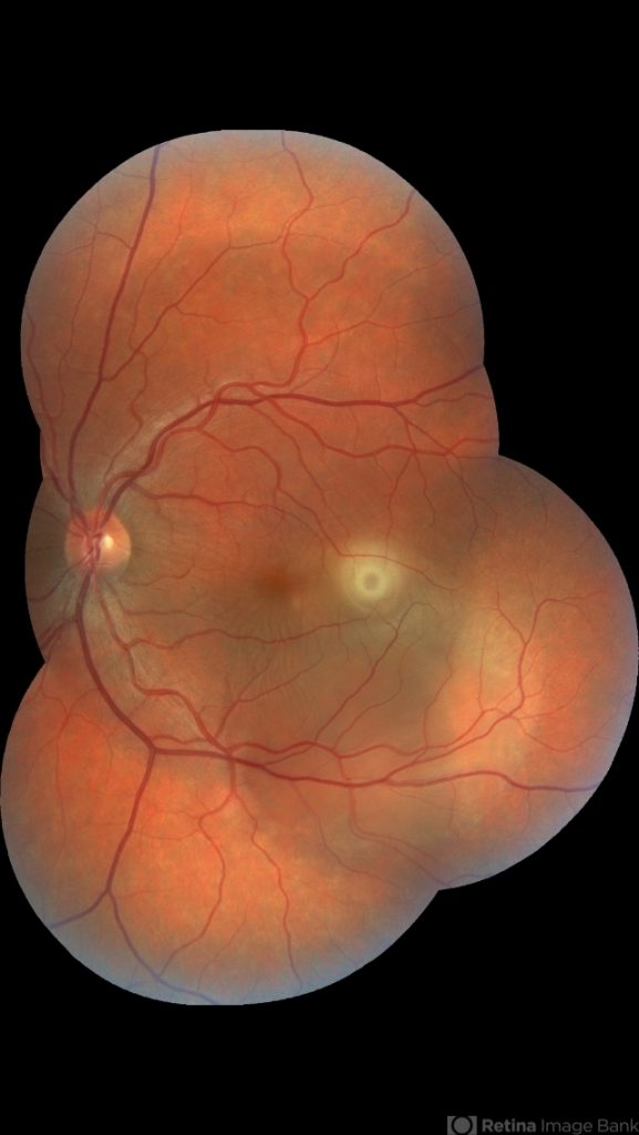
At the time of admission, the best corrected visual acuity was 0.5 in the right eye and 1.0 in the left eye. Based on fundoscopy, the serous detachment had a well-defined margin of 1 disc diameter and was observed in the posterior pole.
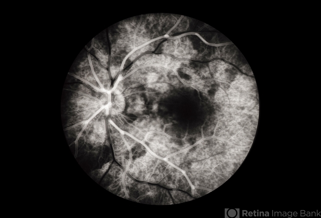
Fluorescein angiography (FAG) identified the point-like hyperfluorescent findings of the early stage. The fluorescent substance was found to slightly spread from the leakage point with time.
There was serous neurosensory detachment on optical coherence tomography. Upon slit lamp examination, there was no evidence of inflammation in the anterior or posterior chamber.
There was a lack of systemic causative factors for this patient. There were also no notable findings upon hematologic assessment performed in the Department of Endocrine Pediatrics. The patient was diagnosed with Central Serous Chorioretinopathy (CSCR).
Central Serous Chorioretinopathy (CSCR) DISEASE entity
Central Serous Chorioretinopathy (CSCR) is the fourth most common retinopathy after age-related macular degeneration, diabetic retinopathy, and branch retinal vein occlusion.
CSCR typically occurs in males in their 20s to 50s who exhibit acute or sub-acute central vision loss or distortion.
Other common complaints include micropsia, metamorphopsia, hyperopic (most common) or myopic shift, central scotoma, and reduced contrast sensitivity and color saturation.
No underlying pathophysiologic mechanisms have been proven, but CSCR is thought to occur due to hyper-permeable choroidal capillaries, which, in association with retinal pigment dysfunction, cause a serous detachment of the neurosensory retina.
Recurrence occurs in about 31% of patients with Central Serous Chorioretinopathy (CSCR), though the recurrence rate has been quoted to be up to 50% in most texts.
The disease was first recognized by Albrecht von Graefe in 1866 and was named central recurrent retinitis. Since then it has been reported under a variety of names such as idiopathic flat detachment of the macula by Walsh et al, central angiospastic retinopathy by Gifford et al, and central serous retinopathy by Straatsma et al. The condition was named Idiopathic central serous chorioretinopathy by Gass et al in 1967.
Most patients who present with CSCR are between the ages of 28 to 68 years with an average age of 43 years. Those who are over 50 years of age are more likely to have bilateral disease (50%) with RPE loss and choroidal neovascularization compared to those less than 50 years of age (28.4%).
Central Serous Chorioretinopathy (CSCR) tends to affect males (9.9/100,000) about six times more than females (1.7/100,000).3 Similar prevalence of CSCR was noted in Caucasian, African American, and Asian populations.
Studies have suggested that there may be a temporal predominance in spring months (March, April, May, and June) but the results were not statistically significant.
Weenink and colleagues studied the family members of 27 patients with bilateral CSCR and found that in 52% of the families, at least one relative was affected.
Of the studied family members, 27.5% suffered from chronic CSCR in at least one eye, suggesting a genetic predisposition to central serous chorioretinopathy.
Most CSC cases are diagnosed in patients with no refractive error or mild hyperopia. Common risk factors, apart from the ones described in “etiology” include pregnancy, antibiotic use, alcohol use, untreated hypertension, and obstructive sleep apnea.
Fok found that patients with psychiatric disorders such as depression are more likely to have a recurrence. Other associations include Cushing syndrome, systemic lupus erythematosus (SLE), organ transplantation, and end-stage renal disease.
Neurosensory detachments and pigment epithelial detachments simulating Central Serous Chorioretinopathy (CSCR) may also be noted with choroidal ischemia in SLE, Goodpasture syndrome, polyarteritis nodosa, thrombotic thrombocytopenic parpura, disseminated intravascular coagulation, granulomatosis with polyangiitis (formerly Wegener granulomatosis), malignant hypertension, and pregnancy-induced hypertension.
Central Serous Chorioretinopathy (CSCR) MANAGEMENT
As CSCR usually resolves spontaneously within 2 to 3 months, observation is currently the standard of care for newly presenting cases.
For chronic CSCR, recurrent CSCR, and acute CSCR in functionally monocular patients, treatment should be discussed.
Risk Factors and Risk Factor Modification
Exogenous steroids of any route (oral, intramuscular, intranasal, etc.) have been strongly linked to an increased risk of Central Serous Chorioretinopathy (CSCR), even at doses as low as 10 mg per day.
Discontinuation of steroids is highly recommended; however, if patients must remain on steroids, reductions in steroid dose have been shown to increase the speed of CSCR resolution.
Because the eye care provider is typically not the person who prescribed these steroids, it is important to maintain close provider-to-provider communication so that the patient’s systemic disease is concurrently managed.
While there are several other risk factors that have been associated with CSCR, modification strategies have not yielded consistent results like discontinuation of exogenous steroids.
Patients with Type A personality should be addressed by lifestyle modification and stress management, although there is an absence of high-quality data to support this. H. pylori has been linked to CSCR; its atherosclerosis-inducing cytotoxins are believed to lead to choroidal vessel damage.

Several studies investigated the effects of screening and treating H. pylori in patients with CSCR but resulted in discordant results on SRF reabsorption and VA improvement.
Similarly, long-standing hypertension has been associated with CSCR; however, treatment with anti-hypertensive agents such as propranolol and metoprolol has yielded no beneficial effects. Eplerenone is not superior to a placebo for improving chronic Central Serous Chorioretinopathy (CSCR) in a clinical trial.
ٌRead more about management strategies
Would you have interest in taking retinal images with your smartphone?
Fundus photography is superior to fundus analysis as it enables intraocular pathologies to be photo-captured and encrypted information to be shared with colleagues and patients.
Recent technologies allow smartphone-based attachments and integrated lens adaptors to transform the smartphone into a portable fundus camera and Retinal imaging by smartphone.
RETINAL IMAGING BY YOUR SMARTPHONE
REFERENCES
- American Academy of Ophthalmology. Central Serous Chorioretinopathy. Basic and Clinical Science Course, Section 12. Retina and Vitreous. San Francisco: American Academy of Ophthalmology; 2022-2023:215-221.
- Porter D, Gregori NZ. Central Serous Chorioretinopathy. American Academy of Ophthalmology.
- Porter D, Vemulakonda GA. Blood Pressure. American Academy of Ophthalmology.
- AAO, Laser Photocoagulation of the Retina and Choroid, 1997, p. 237-240.
- Bousquet E, Beydoun T, Rothschild PR, et al. Spironolactone for Nonresolving Central Serous Chorioretinopathy: A Randomized Controlled Crossover Study. Retina. 2015 May 26.
- Bousquet E, et al. Mineralocorticoid receptor antagonism in the treatment of chronic central serous chorioretinopathy: a pilot study. Retina. 2013 Nov-Dec; 33(10):2096-102.
- Haimovici R, Koh S, Gagnon DR, et al. Risk factors for central serous Chorioretinopathy: a case-control study. Ophthalmology. 2004;111:244-9.
- Jampol LM, Weinreb R, Yannuzzi L. Involvement of corticosteroids and catecholamines in the pathogenesis of central serous Chorioretinopathy: a rationale for new treatment strategies. Ophthalmology. 2002;109:1765-6.
