
Rhegmatogenous retinal detachment (RRD) occurs when liquefied vitreous passes through a full-thickness retinal break and separates the neurosensory retina from the retinal pigment epithelium.
It is the most common type of retinal detachment and one of the few retinal emergencies where the timing of surgery can change the visual outcome.
Most population studies put the incidence at roughly one in 10,000 people per year, with a higher rate in myopic and pseudophakic eyes.
The diagnosis is made by looking for the break, and the surgical choice depends on where it is and what the rest of the eye looks like.
What Is a Rhegmatogenous Retinal Detachment?
The word rhegma means break.
Three conditions must be present for an RRD:
- A retinal break
- Liquefied vitreous that can pass through the break
- Traction or fluid dynamics that keep the fluid flowing
Retinal tears usually develop at the time of acute posterior vitreous detachment, when vitreous traction pulls a flap of retina from its attachment.
Atrophic holes and dialyses can also lead to detachment without traction, and they are more common in younger patients and after trauma.
Risk Factors
The main risk factors are:
- Myopia, especially high myopia
- Lattice degeneration (see lattice degeneration)
- Previous cataract surgery, particularly with posterior capsule rupture
- Previous retinal detachment in the fellow eye
- Family history and inherited vitreoretinopathies such as Stickler syndrome
- Ocular trauma
- Retinal breaks discovered at the time of a symptomatic PVD
A symptomatic horseshoe tear carries a much higher risk of detachment than an asymptomatic round hole (see horseshoe tears and posterior vitreous detachment).
Fundus Explorer Pro
Photograph the retinal findings described here with the phone already in your pocket — 22 D optics and built-in illumination in one handheld unit.
From Choroida — the team behind this siteSymptoms
Patients describe a sudden shower of floaters, flashes of light, and a shadow or curtain that spreads from the periphery.
The shadow starts in the field opposite the detached retina, so a superior detachment causes an inferior field defect.
Central vision is lost when the macula detaches, and acuity falls sharply.
Some detachments, particularly inferior ones or those from atrophic holes, progress slowly and cause few symptoms until the macula is involved.
Examination
Clinical Signs
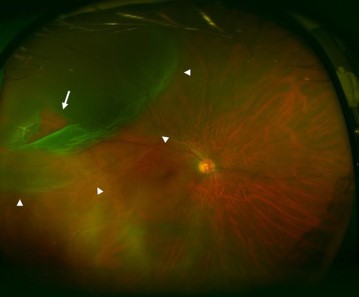
The retina appears gray and corrugated, and it moves with eye movement.
Subretinal fluid follows gravity and posture.
Other findings include:
- Lower intraocular pressure in the affected eye
- Pigment cells in the anterior vitreous (Shafer sign, or tobacco dust)
- A relative afferent pupillary defect when the detachment is extensive
- Visual field defect on confrontation
Locating the Break
Dilated examination with scleral depression and indirect ophthalmoscopy is the standard.
Lincoff’s rules help predict where the primary break lies from the shape of the detachment:
- A superior temporal or superior nasal detachment has its primary break within about one and a half clock hours of the highest border
- A total detachment with a superior wedge has its break at 12 o’clock
- An inferior detachment whose higher side is nasal or temporal has its break on that higher side
- An inferior detachment that is equal on both sides has its break near 6 o’clock
The rules are helpful but they are not a substitute for finding the break, and multiple breaks are common.
B-scan ultrasonography helps when the view is poor, and OCT shows whether the macula is on or off.
Differential Diagnosis
Conditions that can mimic RRD include:
- Degenerative retinoschisis (see senile retinoschisis)
- Choroidal detachment
- Exudative retinal detachment
- Tractional retinal detachment
- Posterior vitreous detachment with vitreous hemorrhage
The distinction matters because exudative and tractional detachments need different management (see retinal detachment lookalikes).
Management
Urgency
Macula-on detachments should be repaired promptly, often within 24 hours, because the macula can detach at any time (see macula-on RRD).
Macula-off detachments are still urgent, although a short delay of a day or two does not appear to worsen the final acuity in some series.
Posturing the patient so that the break lies in the lowest position and the retina is not further detached can help while waiting.
Choosing a Procedure
Options include laser or cryotherapy for retinal breaks without detachment, pneumatic retinopexy, scleral buckling, and pars plana vitrectomy.
- Scleral buckle is favored in young phakic patients with a clear posterior vitreous, without PVD, and with retinal breaks anterior or in the periphery (see scleral buckle surgery)
- Pars plana vitrectomy suits pseudophakic eyes, media opacity, posterior breaks, and cases with PVR or vitreous hemorrhage (see pars plana vitrectomy)
- Pneumatic retinopexy works for a single break or a group of breaks within one clock hour in the superior 8 clock hours
The PIVOT trial compared pneumatic retinopexy with vitrectomy in eyes with a single break or clustered breaks.
Single-surgery success favored vitrectomy, about 93% versus 81%, but visual acuity and metamorphopsia favored pneumatic retinopexy.
The Scleral Buckling versus Primary Vitrectomy (SPR) study compared the two operations directly and remains a useful reference when the choice depends on lens status.
Complications
Proliferative vitreoretinopathy is the main cause of surgical failure (see proliferative vitreoretinopathy).
Other complications include new breaks, cataract, raised IOP, macular pucker, diplopia after buckling, and endophthalmitis.
Prevention and Counseling
Prophylactic treatment of asymptomatic lattice or round holes is not routinely recommended, because the risk of detachment from these lesions is low and laser has its own risks (see laser retinopexy).
Symptomatic breaks, particularly fresh horseshoe tears with persistent traction, are treated with laser or cryotherapy within days.
Patients with risk factors should be told exactly what to watch for: a new shower of floaters, flashes, or a shadow in the field.
They should also know to seek examination the same day if any of these appear, because the window in which the macula is still attached is short.
After cataract surgery in a highly myopic eye, the patient should receive the same advice, and the fellow eye should be examined with scleral depression.
Prognosis
Anatomic success with a single surgery is high in most series.
Final vision depends mainly on macular status and duration of macular detachment, with macula-off eyes often recovering less than macula-on eyes.
Fellow-eye examination is part of every evaluation, and patients should be taught to report new flashes, floaters, or field loss promptly.


Document what you see
Two smartphone imaging tools built for everyday clinic use — one for the slit lamp, one for the fundus.
From Choroida — the team behind this siteReferences
- Steel D. Retinal detachment. BMJ Clin Evid. 2014;2014:0710.
- Mitry D, Charteris DG, Fleck BW, Campbell H, Singh J. The epidemiology of rhegmatogenous retinal detachment: geographical variation and clinical associations. Br J Ophthalmol. 2010;94:678-684.
- Lincoff H, Gieser R. Finding the retinal hole. Arch Ophthalmol. 1971;85:565-569.
- Heimann H, Bartz-Schmidt KU, Bornfeld N, et al. Scleral buckling versus primary vitrectomy in rhegmatogenous retinal detachment: a prospective randomized multicenter clinical study. Ophthalmology. 2007;114:2142-2154.
- Hillier RJ, Felfeli T, Berger AR, et al. The Pneumatic Retinopexy versus Vitrectomy for the Management of Primary Rhegmatogenous Retinal Detachment Outcomes Randomized Trial (PIVOT). Ophthalmology. 2019;126:531-539.
- Kuhn F, Aylward B. Rhegmatogenous retinal detachment: a reappraisal of its pathophysiology and treatment. Ophthalmic Res. 2014;51:15-31.
Test yourself
A few questions straight from this article.
-
What produces a rhegmatogenous retinal detachment?
Rhegma means break: liquefied vitreous passes through a full-thickness retinal break and separates the neurosensory retina from the retinal pigment epithelium. -
What is the approximate annual incidence of rhegmatogenous retinal detachment?
Most population studies put the incidence at roughly one in 10,000 people per year, with higher rates in myopic and pseudophakic eyes. -
By Lincoff's rules, where does the primary break lie in a superior temporal detachment?
Lincoff's rules place the primary break in a superior temporal or superior nasal detachment within roughly one and a half clock hours of the highest border of the detachment. -
A patient with a superior rhegmatogenous retinal detachment reports a field shadow in which position?
The shadow appears in the field opposite the detached retina, so a superior detachment produces an inferior field defect. -
Which finding is known as Shafer sign, or tobacco dust?
Shafer sign is pigment cells visible in the anterior vitreous, one of the supporting signs alongside lower intraocular pressure and a field defect on confrontation. -
Pneumatic retinopexy is suited to which break configuration?
Pneumatic retinopexy works for a single break, or breaks clustered within one clock hour, located in the superior eight clock hours. -
In the PIVOT trial, what was single-surgery success for vitrectomy versus pneumatic retinopexy?
Single-surgery anatomic success favoured vitrectomy at about 93% against 81%, yet visual acuity and metamorphopsia outcomes favoured pneumatic retinopexy. -
Which eye is best suited to scleral buckling for rhegmatogenous retinal detachment?
Buckling is favoured in young phakic eyes with a clear posterior vitreous and anterior or peripheral breaks, while vitrectomy suits pseudophakic eyes, media opacity and posterior breaks. -
What is the main cause of surgical failure after rhegmatogenous retinal detachment repair?
Proliferative vitreoretinopathy is the leading cause of failure; new breaks, cataract, raised pressure, macular pucker and diplopia are other recognised complications. -
How quickly should a macula-on rhegmatogenous retinal detachment be repaired?
Macula-on detachments warrant repair promptly, often within 24 hours, because the macula can detach at any moment; macula-off eyes are still urgent.

Writes and reviews the clinical articles on Educate. Every post is checked against peer-reviewed sources before it goes live.


