
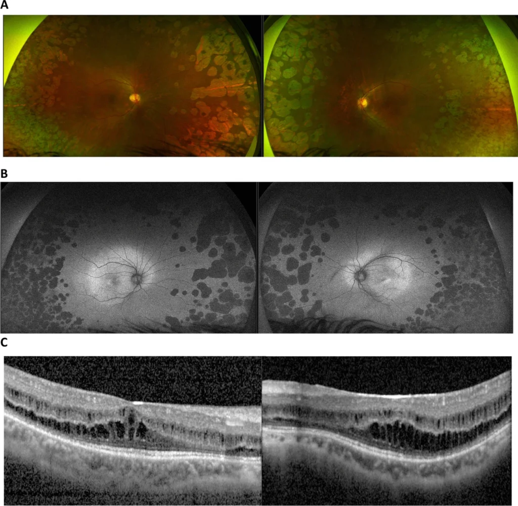
Fundus autofluorescence (FAF) images the natural fluorescence of lipofuscin and other fluorophores within the retinal pigment epithelium, producing a map of RPE health that requires no dye and no injection.
Where a fundus photograph shows what the retina looks like, autofluorescence shows something closer to how metabolically stressed or depleted the RPE actually is at each point, which is information a colour photograph alone cannot provide.
How Fundus Autofluorescence Works
Lipofuscin accumulates within RPE cells as a natural by-product of the RPE’s ongoing task of processing shed photoreceptor outer segments, and it fluoresces when excited by light of an appropriate wavelength, typically blue or near-infrared depending on the system used.
A specific excitation wavelength is used to stimulate this fluorescence, and the emitted light is captured to build an image, entirely without any exogenous dye or intravenous injection.
The resulting signal reflects RPE cell activity and lipofuscin content rather than blood flow, which is the fundamental difference from angiography, and is what makes autofluorescence so useful for tracking degenerative and atrophic disease specifically.
Interpreting the Signal
Hyperautofluorescence
Increased signal reflects excess lipofuscin accumulation within stressed or dysfunctional RPE cells, seen in conditions including some pattern dystrophies, drusen, and the junctional zone at the margin of areas of geographic atrophy, where RPE cells are under metabolic stress before they die.
Hypoautofluorescence
Reduced or absent signal indicates loss of RPE cells and their lipofuscin content, most classically seen as the sharply demarcated dark patches of geographic atrophy, but also in areas of chorioretinal scarring, dense haemorrhage blocking the underlying signal, or RPE atrophy from other degenerative and inherited retinal diseases.
Fundus Explorer Pro
Photograph the retinal findings described here with the phone already in your pocket — 22 D optics and built-in illumination in one handheld unit.
From Choroida — the team behind this siteClinical Applications
Age-Related Macular Degeneration
Autofluorescence precisely delineates the borders of geographic atrophy, often more clearly than colour photography, and the hyperautofluorescent junctional zone surrounding an atrophic lesion is used as a marker of RPE tissue at risk of imminent progression.
Inherited Retinal Disease

Conditions such as Stargardt disease, retinitis pigmentosa, and gyrate atrophy show characteristic autofluorescence patterns that often reveal the true extent of RPE involvement well beyond what is visible on colour fundus photography alone, and serial imaging over time is used to track disease progression in these conditions.
Central Serous Chorioretinopathy
Autofluorescence can help distinguish acute from chronic disease and outline the extent of RPE change from long-standing or recurrent subretinal fluid, complementing OCT findings.
Monitoring Drug Toxicity
Hydroxychloroquine retinopathy produces a characteristic pattern of parafoveal or, in some populations, more peripheral autofluorescence change, and FAF is one of the recommended screening modalities used alongside visual fields and OCT for patients on long-term treatment.
Advantages Over Other Imaging
Being entirely non-invasive with no dye or injection required, autofluorescence can be repeated as often as needed for monitoring, without the allergic reaction risk or contraindications relevant to fluorescein or indocyanine green angiography.
It is fast, typically taking only seconds to acquire, and reveals RPE-level pathology that is often not obvious, or not obvious to the same degree, on standard colour fundus photography.
Limitations
Media opacity, including cataract or vitreous haemorrhage, degrades image quality and can obscure the underlying autofluorescence signal.
Autofluorescence does not show vascular flow or leakage, so it cannot replace fluorescein angiography when the clinical question is specifically about active leakage or neovascularisation rather than RPE status.
Interpretation requires correlation with other imaging and clinical findings, since hyperautofluorescence and hypoautofluorescence each have multiple possible causes, and the pattern alone does not always point to a single diagnosis without additional clinical context.
Clinical Impact
Autofluorescence has become a standard part of monitoring geographic atrophy, including in clinical trials of complement inhibitor therapy, where precise measurement of lesion size and growth rate over time depends heavily on this imaging modality.
In inherited retinal disease, it contributes meaningfully to diagnosis, prognostic counselling, and increasingly to trial eligibility assessment as gene therapy and other treatments reach clinical use for specific conditions.
As a non-invasive, repeatable, and information-rich technique, autofluorescence has moved from a research tool to a routine part of retinal imaging across a wide range of conditions, sitting alongside OCT as a standard component of a comprehensive retinal imaging workup.


Document what you see
Two smartphone imaging tools built for everyday clinic use — one for the slit lamp, one for the fundus.
From Choroida — the team behind this siteReferences
- Schmitz-Valckenberg S, Holz FG, Bird AC, Spaide RF. Fundus autofluorescence imaging: review and perspectives. Retina. 2008.
- Holz FG, Bindewald-Wittich A, Fleckenstein M, et al. Progression of geographic atrophy and impact of fundus autofluorescence patterns in age-related macular degeneration. American Journal of Ophthalmology. 2007.
- Marmor MF, Kellner U, Lai TY, et al. Recommendations on screening for chloroquine and hydroxychloroquine retinopathy. Ophthalmology. 2016.
- Fundus Autofluorescence. EyeWiki, American Academy of Ophthalmology.
- Fundus Autofluorescence Imaging. StatPearls, NCBI Bookshelf.
Test yourself
A few questions straight from this article.
-
Which molecule is the principal source of the fundus autofluorescence signal?
Fundus autofluorescence images the natural fluorescence of lipofuscin and related fluorophores inside RPE cells, mapping RPE health without any dye. -
Lipofuscin accumulates inside retinal pigment epithelial cells as a by-product of which process?
Lipofuscin builds up as a natural by-product of the RPE's continuous task of processing shed photoreceptor outer segments. -
What practical advantage does fundus autofluorescence hold over dye-based angiography?
Because it needs no exogenous dye, autofluorescence can be repeated as often as monitoring requires, avoiding the allergic risk and contraindications of fluorescein or ICG. -
The fundus autofluorescence signal primarily reflects which property of the retina?
The signal reports RPE activity and lipofuscin content rather than blood flow, which is the fundamental difference from angiography and why it suits atrophic disease. -
What does hyperautofluorescence indicate about the retinal pigment epithelium?
Increased signal reflects lipofuscin accumulating in stressed or dysfunctional RPE cells, as seen in pattern dystrophies, drusen and the margins of geographic atrophy. -
Sharply demarcated dark patches of hypoautofluorescence most classically represent which lesion?
Reduced or absent signal means loss of RPE cells and their lipofuscin, seen most classically as the crisply bordered dark patches of geographic atrophy. -
What does the hyperautofluorescent junctional zone surrounding geographic atrophy signify?
The hyperautofluorescent rim marks metabolically stressed RPE bordering the atrophic lesion, and serves as a marker of tissue likely to be lost next. -
For hydroxychloroquine retinopathy screening, fundus autofluorescence is recommended alongside which tests?
Hydroxychloroquine retinopathy produces a characteristic parafoveal, or in some populations more peripheral, autofluorescence change, and FAF is used with visual fields and OCT for screening. -
When the clinical question is active leakage or neovascularisation, which investigation remains necessary?
Autofluorescence shows no vascular flow or leakage, so it cannot substitute for fluorescein angiography when leakage or neovascularisation is the question. -
In Stargardt disease and retinitis pigmentosa, what does autofluorescence characteristically demonstrate?
These inherited retinal diseases show characteristic patterns that reveal the true extent of RPE involvement well beyond what colour fundus photography shows, and serial imaging tracks progression.

Writes and reviews the clinical articles on Educate. Every post is checked against peer-reviewed sources before it goes live.


