
Optic disc pit maculopathy is a serous and schisis-like detachment of the macula that develops in eyes with a congenital optic disc pit, and it stands out among causes of macular fluid because there is no retinal break to find, no matter how carefully the surgeon looks.
The absence of a break has fueled decades of debate about where the fluid actually comes from, and that debate still shapes how the condition is treated.
What Is an Optic Disc Pit?
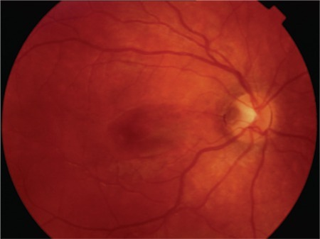
A congenital optic disc pit is a focal excavation of the optic nerve head, usually located at the temporal or inferotemporal aspect of the disc, present from birth and typically an incidental finding unless maculopathy develops.
A minority of eyes with a disc pit, roughly a quarter to a half in various series, go on to develop maculopathy, usually in young adulthood, causing metamorphopsia and reduced central vision (see optic pits).
Where Does the Fluid Come From?
Several sources have been proposed, and the true mechanism may involve more than one.
- Cerebrospinal fluid tracking from the subarachnoid space through the pit into the retina, given the pit’s anatomic connection to the optic nerve sheath in some eyes
- Liquefied vitreous entering through the pit, driven by vitreous traction at the disc margin
- Fluid derived from leaky vessels at the base of the pit itself
OCT has shown that the fluid typically first accumulates as a schisis-like separation within the inner retinal layers, connecting to the pit, before a secondary outer layer serous detachment develops, which helps explain the layered, complex appearance often seen on imaging.
Fundus Explorer Pro
Photograph the retinal findings described here with the phone already in your pocket — 22 D optics and built-in illumination in one handheld unit.
From Choroida — the team behind this siteClinical and Imaging Features
- Reduced central vision and metamorphopsia, typically presenting in the third or fourth decade of life
- A visual field defect corresponding to the disc pit’s location can be present even without maculopathy, from the structural nerve fiber layer disruption at the pit itself
- OCT shows an inner retinal schisis-like cavity connecting the disc pit to the macula, often with an outer layer serous detachment, sometimes with a macular hole developing in longstanding cases
- Fluorescein angiography typically shows little to no leakage, which helps distinguish this from other causes of macular edema and supports a mechanical rather than inflammatory or vascular process
Differential Diagnosis
- Central serous chorioretinopathy, which shows angiographic leakage and choroidal thickening, absent in optic disc pit maculopathy
- Rhegmatogenous retinal detachment, excluded by the absence of a peripheral retinal break
- Optic disc coloboma with associated retinal detachment, a related but distinct congenital anomaly with a larger excavation
- X-linked retinoschisis, which has a different, typically bilateral, spoke-wheel macular pattern and family history
Management
Observation
Some cases are observed initially, particularly with mild or stable visual symptoms, since spontaneous improvement occurs in a minority of eyes.
Laser Photocoagulation
Laser applied to the temporal disc margin, between the pit and the macula, aims to create a chorioretinal adhesion that blocks further fluid tracking from the pit into the macula, though its effectiveness is inconsistent across reported series.
Pars Plana Vitrectomy
Vitrectomy with induction of a posterior vitreous detachment, often combined with laser to the disc margin and gas tamponade, is the most commonly used treatment for vision-threatening maculopathy, aiming to relieve vitreous traction at the disc that is thought to drive fluid into the pit.
Internal limiting membrane peeling is used by some surgeons as an adjunct, based on the idea that residual traction from the ILM contributes to persistent or recurrent fluid.
Inner Fenestration and Other Techniques
Some surgeons create a small fenestration or “drainage” opening in the inner retinal schisis wall at the macula to allow trapped fluid to escape into the vitreous cavity, an approach used particularly for eyes with a large schisis cavity.
Prognosis
Anatomic improvement is achieved in a majority of surgically treated eyes, though visual recovery is often incomplete, particularly when maculopathy has been long-standing or a macular hole has developed.
Because the natural history is variable and some eyes stabilize without surgery, the decision to operate weighs the degree of visual impairment and symptom duration against the uncertain but real possibility of surgical benefit.


Document what you see
Two smartphone imaging tools built for everyday clinic use — one for the slit lamp, one for the fundus.
From Choroida — the team behind this siteReferences
- Sobol WM, Blodi CF, Folk JC, Weingeist TA. Long-term visual outcome in patients with optic nerve pit and serous retinal detachment of the macula. Ophthalmology. 1990;97:1539-1542.
- Lincoff H, Kreissig I. Optical coherence tomography of pneumatic displacement of optic disc pit maculopathy. Br J Ophthalmol. 1998;82:367-372.
- Hirakata A, Inoue M, Hiraoka T, McCuen BW 2nd. Vitrectomy without laser treatment or gas tamponade for macular detachment associated with an optic disc pit. Ophthalmology. 2012;119:810-818.
- Ooto S, Mittra RA, Ridley ME, Spaide RF. Vitrectomy with inner retinal fenestration for optic disc pit maculopathy. Ophthalmology. 2014;121:1727-1733.
Test yourself
A few questions straight from this article.
-
What distinguishes optic disc pit maculopathy from most other causes of macular detachment?
The macular fluid develops without any retinal break, however carefully the surgeon looks, which is what sets this entity apart. -
A congenital optic disc pit is usually located at which part of the disc?
The pit is a focal excavation of the nerve head, usually sited temporally or inferotemporally, and present from birth. -
Which has been proposed as a source of the fluid in optic disc pit maculopathy?
Cerebrospinal fluid from the subarachnoid space is one proposed source, alongside liquefied vitreous and leaky vessels at the base of the pit. -
On OCT, how does fluid in optic disc pit maculopathy typically first appear?
Fluid first collects as a schisis-like separation within the inner retinal layers connecting to the pit, before a secondary outer layer serous detachment develops. -
What does fluorescein angiography typically show in optic disc pit maculopathy?
The relative absence of leakage separates this from other causes of macular oedema and supports a mechanical rather than inflammatory or vascular process. -
Which features of central serous chorioretinopathy are absent in optic disc pit maculopathy?
Central serous chorioretinopathy shows angiographic leakage and a thickened choroid, neither of which is found in optic disc pit maculopathy. -
A visual field defect can occur with an optic disc pit even without maculopathy. Why?
Structural nerve fibre layer disruption at the pit produces a field defect corresponding to the pit's location, independent of any macular fluid. -
What is laser photocoagulation at the temporal disc margin intended to achieve?
Laser between the pit and the macula aims to create a chorioretinal adhesion that blocks further fluid tracking, though reported effectiveness is inconsistent. -
Which treatment is most commonly used for vision-threatening optic disc pit maculopathy?
Vitrectomy with posterior vitreous detachment induction, often with laser to the disc margin and gas tamponade, relieves the vitreous traction thought to drive fluid into the pit. -
What is the usual outcome after surgery for optic disc pit maculopathy?
Most surgically treated eyes improve anatomically, but visual recovery is frequently partial, especially with longstanding disease or a macular hole.

Writes and reviews the clinical articles on Educate. Every post is checked against peer-reviewed sources before it goes live.


