
Siegrist streaks are the long-term scar of acute choroidal ischemia in severe hypertension.
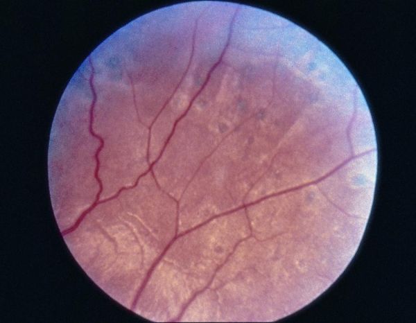
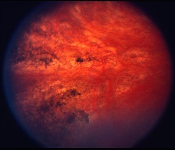
They are thin, linear, hyperpigmented lines that run along choroidal vessels, often in the posterior pole, and they tell you that the patient once had a serious hypertensive crisis even if nobody has recorded it.
How they form
In accelerated or malignant hypertension, choroidal arterioles are acutely occluded.
The overlying RPE dies, leaving a pale yellow patch, an Elschnig spot.
As the weeks pass, RPE cells proliferate and hyperpigment at the borders, and the lesion matures into a pigmented spot.
When a linear vessel is involved, the same process leaves a line, and that line is a Siegrist streak (see hypertensive choroidopathy).
Appearance
- Linear, dark, sometimes branching streaks, following the course of a choroidal vessel
- Often paired with scattered round pigmented Elschnig scars
- Located chiefly in the posterior pole and mid-periphery
- Usually bilateral and asymmetric
Fundus Explorer Pro
Photograph the retinal findings described here with the phone already in your pocket — 22 D optics and built-in illumination in one handheld unit.
From Choroida — the team behind this siteSettings in which they occur
- Malignant hypertension, in patients who survived the acute phase
- Pre-eclampsia and eclampsia, where choroidal changes are part of the acute presentation
- Renal disease with severe hypertension
- Other choroidal vasculopathies such as thrombotic microangiopathy and some vasculitides
Clinical significance
Streaks themselves rarely impair vision unless they involve the fovea.
They are best seen as a sign, evidence that blood pressure was once high enough to damage the choroid.
In a young woman with a fundus like this, a history of eclampsia is worth asking about.
Investigation
- Blood pressure measurement and a cardiovascular and renal review
- Fluorescein angiography, which shows window defects and patchy filling delay
- OCT, which may demonstrate RPE irregularity and sometimes thin choroid under the streaks
The acute stages that precede them
The sequence is worth knowing because it explains the findings in front of you.
In the acute phase of a hypertensive crisis, fluorescein angiography shows delayed or patchy choroidal filling because the choriocapillaris lobules are occluded.
The RPE overlying those lobules becomes edematous, and serous retinal detachments can form, producing visual blur.
As the pressure is controlled, fluid resolves, and RPE atrophy leaves a mottled window defect.
Months later, pigment migrates to the edges and a pigmented spot or streak is the end result.
Because the retinal vessels themselves can look normal in young patients who develop acute severe hypertension, choroidal changes sometimes offer the only clue that the pressure was dangerously high.
Who to think of
- Young women after pre-eclampsia, especially if blurred vision during pregnancy was dismissed
- Patients with renal failure and poorly controlled blood pressure
- Patients with scleroderma renal crisis or thrombotic microangiopathy
- Anyone with an unexplained pigmented pattern in the posterior pole and a history of hypertension
Management
There is no treatment of the streaks.
Management is control of the underlying disease, with attention to renal function, and monitoring the eye for associated retinal vascular events.
What the acute lesions look like
In the first days of a hypertensive crisis, the choroid shows patchy filling delay, and the retina may develop serous detachments over the affected areas. The pale yellow patches at the RPE level, the Elschnig spots, may be easy to miss unless the pupil is dilated, and fluorescein shows them as areas of delayed filling with later leakage. Streaks and pigmented spots appear weeks later as the RPE heals.
Why the old lesions are worth recognizing
A patient with Siegrist streaks and a normal blood pressure today may have had malignant hypertension years earlier, perhaps in pregnancy, and may now have renal damage or a risk of recurrent hypertension. Asking about previous eclampsia, kidney disease, or hospital admissions for high pressure can reveal an important history that was never connected with the eye.
In a young person with unexplained pigmented streaks, request a blood pressure check, renal function, and urinalysis. Document the lesions photographically, and mention them in any letter to the physician, because they are objective evidence of past severe hypertension.
Pitfalls
- Do not attribute linear pigmentation to myopia or old trauma without asking about blood pressure
- Do not forget that choroidal ischemia can occur in the absence of retinal vascular changes, especially in younger patients
- Do not rely on a normal blood pressure at the visit to exclude past severe hypertension
- Remember that similar changes can follow thrombotic microangiopathy, lupus, and some vasculitides, so a systemic review is worthwhile
Documenting the finding
Describe the number, distribution, and orientation of the streaks, take color and autofluorescence images, and record the history that explains them. This record helps if a later episode of hypertension occurs and helps other clinicians interpret the fundus.


Document what you see
Two smartphone imaging tools built for everyday clinic use — one for the slit lamp, one for the fundus.
From Choroida — the team behind this siteReferences
- Tso MO, Jampol LM. Pathophysiology of hypertensive retinopathy. Ophthalmology. 1982;89:1132-1145.
- Kishi S, Tso MO, Hayreh SS. Fundus lesions in malignant hypertension. I. A pathologic study of experimental hypertensive choroidopathy. Arch Ophthalmol. 1985;103:1189-1197.
- Hayreh SS, Servais GE, Virdi PS. Fundus lesions in malignant hypertension. V. Hypertensive optic neuropathy. Ophthalmology. 1986;93:74-87.
- Valluri S, Adelberg DA, Curtis RS, Olk RJ. Diagnostic indocyanine green angiography in preeclampsia. Am J Ophthalmol. 1996;122:672-677.
