
Panretinal photocoagulation stopped a hundred thousand eyes from going blind long before anyone knew the molecule responsible.
That molecule turned out to be vascular endothelial growth factor, and blocking it directly has since become the single most consequential advance in modern retinal treatment.
Anti-VEGF therapy now underpins the management of the three commonest sight-threatening retinal vascular diseases: wet macular degeneration, diabetic retinopathy and retinal vein occlusion.
Five agents are now in clinical use, each with a different molecular target and a different durability profile.
Choosing between them, and choosing how often to inject, has become one of the highest-stakes decisions in outpatient ophthalmology.
What Is Anti-VEGF Therapy?
Anti-VEGF therapy is treatment that neutralises vascular endothelial growth factor, the principal signalling molecule driving pathological angiogenesis and vascular permeability in the retina.
The agents in current use are:
- Bevacizumab – a full-length antibody developed for oncology and used off-label intravitreally; low cost and widely used where approved formulations are unaffordable
- Ranibizumab – an antibody fragment specifically developed and licensed for intraocular use
- Aflibercept – a fusion protein “trap” binding VEGF-A, VEGF-B and placental growth factor with high affinity, allowing longer dosing intervals
- Brolucizumab – a small single-chain antibody fragment permitting high molar dosing, associated with a distinct intraocular inflammation risk
- Faricimab – a bispecific antibody blocking both VEGF-A and angiopoietin-2, the newest agent and the first to target vessel stability as well as VEGF
The differences between these molecules are not academic – they translate directly into how long a given patient can safely go between injections.
Mechanism of Action
All disease processes treated with anti-VEGF share a common final pathway that the drug class interrupts.
- Retinal ischaemia or RPE dysfunction upregulates VEGF secretion
- VEGF binds its receptors on vascular endothelium, driving new vessel growth, breakdown of tight junctions and vascular leakage
- Anti-VEGF agents bind circulating and tissue VEGF, preventing receptor activation
- The result is reduced vascular permeability, regression of much of the abnormal neovascular complex, and resolution of oedema
- Angiopoietin-2, blocked additionally by faricimab, independently destabilises vessels and amplifies VEGF’s effects – blocking both pathways together produces more durable vessel stabilisation than blocking VEGF alone
Suppression, not cure, is the operative word – VEGF drive typically returns once drug levels fall, which is the basis for ongoing treatment.
Fundus Explorer Pro
Photograph the retinal findings described here with the phone already in your pocket — 22 D optics and built-in illumination in one handheld unit.
From Choroida — the team behind this siteIndications
Established
- Neovascular age-related macular degeneration
- Diabetic macular oedema
- Macular oedema secondary to branch and central retinal vein occlusion
- Myopic choroidal neovascularisation
Expanding
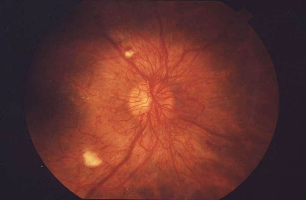
- Proliferative diabetic retinopathy, as an adjunct or alternative to panretinal photocoagulation in selected patients
- Retinopathy of prematurity, now a guideline-supported option alongside laser in appropriate infants
- Neovascular glaucoma, used to regress iris and angle neovascularisation before or alongside definitive glaucoma treatment
The expansion into proliferative diabetic retinopathy is a genuine shift from laser as the default – it is not simply a rescue therapy any more.
Clinical Evidence
- ANCHOR and MARINA established ranibizumab’s superiority over prior standard care in neovascular AMD
- VIEW 1 and VIEW 2 established aflibercept’s non-inferiority to ranibizumab with a longer dosing interval
- Protocol T, run by the DRCR Retina Network, directly compared aflibercept, bevacizumab and ranibizumab in diabetic macular oedema and found aflibercept had an advantage in eyes with worse baseline vision
- TENAYA and LUCERNE demonstrated faricimab could match aflibercept’s efficacy with substantially extended treatment intervals in many patients
- CATT and IVAN found bevacizumab broadly comparable to ranibizumab in AMD, supporting its continued off-label use where cost is a barrier
Protocol T remains particularly influential because it was designed and funded independently of any single manufacturer, giving its head-to-head comparisons unusual credibility.
Treatment Strategy
Loading and Maintenance
- Most regimens begin with monthly loading injections – typically three to five – to achieve maximal anatomical control before spacing treatment out
- Maintenance then follows a fixed, PRN or treat-and-extend schedule depending on the disease and the agent’s durability
Selecting an Agent
- Durability needs – a patient struggling to attend frequent visits benefits from a longer-acting agent
- Disease severity – Protocol T’s subgroup findings still inform first-line choice in diabetic macular oedema with poor baseline vision
- Cost and access – bevacizumab remains the dominant choice in many health systems purely on cost grounds, with comparable efficacy
- Inflammation risk – brolucizumab’s association with retinal vasculitis has narrowed its use in practice since post-marketing data emerged
No single agent is correct for every patient – the choice is a genuine clinical trade-off between durability, cost and individual risk profile.
Safety Considerations
- Endophthalmitis remains the most feared acute complication of the injection procedure itself, not the drug
- Intraocular inflammation, including retinal vasculitis, is a recognised class effect with a higher reported incidence for brolucizumab
- Systemic arterial thromboembolic events are a theoretical concern from systemic VEGF suppression; large pooled analyses have not shown a clear excess risk, but caution is exercised in patients with recent stroke or myocardial infarction
- Geographic atrophy progression with long-term anti-VEGF exposure has been reported in some cohort studies of AMD, though whether this reflects the drug or the natural history of treated eyes remains actively debated
The safety conversation with patients should distinguish clearly between injection-related risk, which is procedural, and drug-related risk, which is pharmacological – conflating the two undermines informed consent.
Prognosis
Anti-VEGF therapy has transformed the natural history of every disease it is used to treat.
- Neovascular AMD, once reliably progressive to legal blindness, now stabilises or improves in the large majority of eyes treated promptly and consistently
- Diabetic macular oedema shows sustained improvement in visual acuity with regular treatment, exceeding what laser alone achieved
- Vein occlusion-related oedema responds well, though the underlying occlusion and ischaemic burden still shape the long-term ceiling on vision
- Real-world outcomes consistently trail trial outcomes, and the gap is explained almost entirely by under-treatment relative to trial protocols
The technology has outpaced the delivery systems built to support it – the next gains in this field are as likely to come from better adherence and longer-acting agents as from a fundamentally new mechanism.


Document what you see
Two smartphone imaging tools built for everyday clinic use — one for the slit lamp, one for the fundus.
From Choroida — the team behind this siteReferences
- Wells JA, Glassman AR, Ayala AR, et al. Aflibercept, bevacizumab, or ranibizumab for diabetic macular edema (Protocol T). New England Journal of Medicine. 2015.
- Heier JS, Khanani AM, Quezada Ruiz C, et al. Efficacy, durability, and safety of faricimab up to every 16 weeks: TENAYA and LUCERNE. The Lancet. 2022.
- CATT Research Group. Ranibizumab and bevacizumab for neovascular age-related macular degeneration. New England Journal of Medicine. 2011.
- Anti-VEGF Agents. EyeWiki, American Academy of Ophthalmology.
- Anti-VEGF Therapy. StatPearls, NCBI Bookshelf.
Test yourself
A few questions straight from this article.
-
What does vascular endothelial growth factor (VEGF) do in the diseased retina?
VEGF is the principal signalling molecule driving new vessel growth, breakdown of tight junctions and leakage, and anti-VEGF agents bind it to prevent receptor activation. -
Which molecules does the fusion protein aflibercept bind?
Aflibercept is a fusion protein trap binding VEGF-A, VEGF-B and placental growth factor with high affinity, which allows longer dosing intervals. -
What distinguishes bevacizumab from the other anti-VEGF agents in retinal practice?
Bevacizumab is a full-length antibody developed for oncology and used off-label intravitreally, and it remains dominant in many health systems purely on cost grounds. -
What role does angiopoietin-2 play in retinal vascular disease?
Angiopoietin-2 independently destabilises vessels and amplifies VEGF's effects, so blocking both pathways produces more durable vessel stabilisation than blocking VEGF alone. -
Why do patients on anti-VEGF therapy generally require ongoing treatment?
Anti-VEGF therapy suppresses an active disease process; VEGF drive typically returns once drug levels fall, which is the basis for continued treatment. -
What did Protocol T find when comparing anti-VEGF agents in diabetic macular oedema?
Protocol T compared aflibercept, bevacizumab and ranibizumab head to head and found an aflibercept advantage in eyes with worse baseline vision, a finding that still guides first-line choice. -
How many monthly loading injections do most anti-VEGF regimens begin with?
Most regimens start with three to five monthly loading injections to achieve maximal anatomical control before treatment is spaced out into a maintenance schedule. -
Which safety signal has narrowed the practical use of brolucizumab?
Intraocular inflammation is a class effect but is reported more often with brolucizumab, and its association with retinal vasculitis has narrowed its use since post-marketing data emerged. -
In which paediatric retinal condition is anti-VEGF now a guideline-supported option alongside laser?
Retinopathy of prematurity is one of the expanding indications, where anti-VEGF is now a guideline-supported option alongside laser in appropriate infants. -
What explains the gap between real-world and clinical trial outcomes of anti-VEGF therapy?
Real-world outcomes consistently trail trial outcomes, and the gap is explained almost entirely by under-treatment compared with trial dosing protocols.

Writes and reviews the clinical articles on Educate. Every post is checked against peer-reviewed sources before it goes live.


