
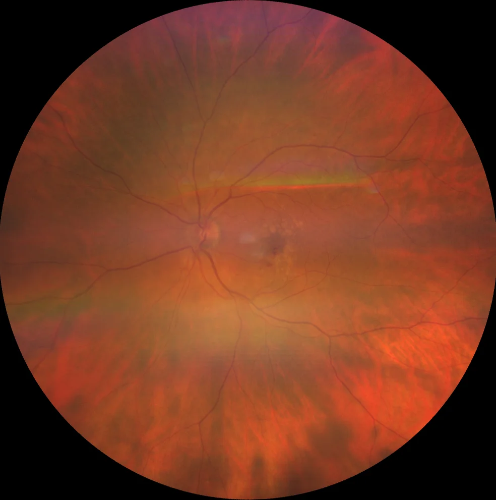
Macular neovascularization (MNV) is the current consensus term for abnormal vessel growth in the macula that originates from the choroid or the retina.
It replaces the older term choroidal neovascularization (CNV), because not all lesions arise from the choroid.
The three types differ in location, imaging, natural history, and response to treatment, so identifying the type helps predict behavior and plan follow-up.
What Is Macular Neovascularization?
Historically, neovascular AMD was described as classic, occult, or predominantly classic based on fluorescein angiography.
OCT and OCT angiography show the actual anatomic location of the vessels, and this proved more informative.
In 2020 a consensus group proposed a nomenclature in which MNV is classified as type 1, type 2, or type 3 according to the relationship of the lesion to the retinal pigment epithelium.
The term neovascular AMD refers to MNV in the setting of AMD, and MNV can also occur in many other diseases.
The Three Types
Type 1: Sub-RPE
Type 1 MNV grows between the RPE and Bruch’s membrane.
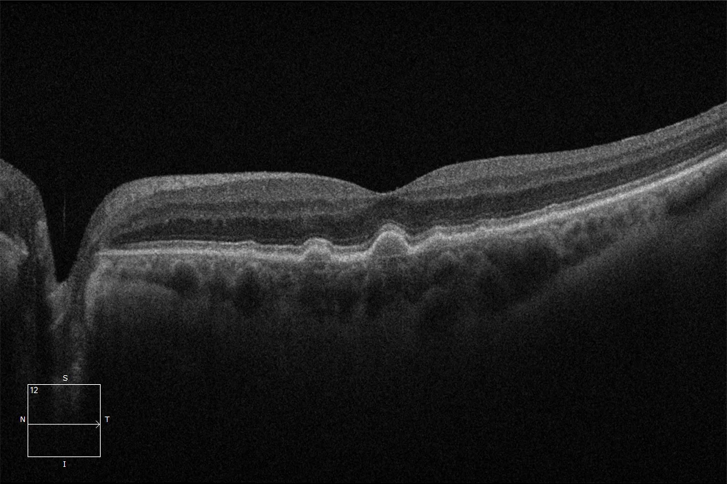
On OCT it appears as a shallow irregular RPE elevation, often with a double-layer sign, and there may be fibrovascular pigment epithelial detachment.
Fluorescein angiography shows stippled or late leakage of an undetermined source, which corresponds to the older occult pattern.
OCT angiography shows a vascular network beneath the RPE.
Type 1 lesions are the most common subtype in patients of European ancestry.
Polypoidal choroidal vasculopathy is regarded as a form of type 1 MNV with aneurysmal dilations (see polypoidal choroidal vasculopathy).
Pachychoroid neovasculopathy is a type 1 lesion arising in eyes with thick choroid (see pachychoroid neovasculopathy).
Type 2: Subretinal
Type 2 MNV grows through the RPE into the subretinal space.
Fluorescein angiography shows well-defined early hyperfluorescence with leakage, the classic pattern.
OCT shows subretinal hyperreflective material above the RPE, often with subretinal fluid and hemorrhage.
Type 2 lesions are more common in younger patients and in non-AMD diseases such as myopia, angioid streaks, and inflammatory conditions.
Type 3: Intraretinal
Type 3 MNV originates in the retinal circulation, usually the deep capillary plexus, and grows toward the RPE.
It was previously called retinal angiomatous proliferation (see retinal angiomatous proliferation).
Features include:
- Intraretinal hyperreflective foci with cystoid edema
- A retinal-retinal or retinal-choroidal anastomosis
- Association with reticular pseudodrusen (see reticular pseudodrusen)
- A tendency to be bilateral, and to occur in elderly women
Type 3 lesions respond to anti-VEGF therapy, though they may require frequent injections and are prone to progression to atrophy.
Mixed Lesions
Types 1 and 2 often coexist, and OCT angiography helps show both components.
Fundus Explorer Pro
Photograph the retinal findings described here with the phone already in your pocket — 22 D optics and built-in illumination in one handheld unit.
From Choroida — the team behind this siteImaging Recommendations
Multimodal imaging is used to type the lesion.
- OCT shows fluid, hyperreflective material, and pigment epithelial detachment
- OCT angiography shows the neovascular network without dye, and it distinguishes sub-RPE from subretinal vessels (see OCT angiography)
- Fluorescein angiography identifies leakage and helps when OCT angiography is limited by hemorrhage
- ICGA shows polyps and branching vascular networks in polypoidal disease
- Autofluorescence shows atrophy and guides prognosis
A single test may not be enough, and the diagnosis usually rests on the combination.
Causes Beyond Age-Related Macular Degeneration
MNV occurs in many conditions.
- Pathologic myopia (see myopic CNV)
- Angioid streaks (see angioid streaks)
- Presumed ocular histoplasmosis (see presumed ocular histoplasmosis)
- Inflammatory choroidal disease such as multifocal choroiditis and punctate inner choroidopathy
- Choroidal rupture after trauma
- Central serous chorioretinopathy
- Macular dystrophies such as vitelliform disease
- Choroidal tumors such as osteoma
The underlying disease influences the natural history and treatment.
Younger patients with MNV should be examined for a secondary cause.
Management
Anti-VEGF injections are first-line therapy for all types.
Regimens include monthly dosing, treat-and-extend, and PRN, and the goal is a dry macula with the fewest injections (see anti-VEGF therapy).
Photodynamic therapy combined with anti-VEGF is used in polypoidal lesions.
Type 2 lesions often respond rapidly, and type 3 may need frequent treatment.
Complications include RPE tears (see RPE tear), subretinal fibrosis, hemorrhage, and macular atrophy.
Patients should understand that the aim is to preserve vision and that treatment is long-term.
Clinical Points by Type
- Type 1: often quiet for a long time, with a slowly enlarging pigment epithelial detachment; a vascularized PED with new fluid should be treated even when acuity is good
- Type 2: typically produces symptoms early because the lesion lies in front of the RPE; visual gains after anti-VEGF are usually greatest in this group
- Type 3: intraretinal cystoid spaces and small hemorrhages near a drusenoid PED are early signs; frequent review is needed because atrophy tends to follow
- Mixed and polypoidal disease: consider ICGA when the response to anti-VEGF is poor or a notched serous PED is seen
Fellow eyes need attention as well.
The risk of MNV in the second eye is substantial in patients with neovascular AMD, and home monitoring plus scheduled OCT allows early treatment.
Follow-Up After Treatment
OCT at every visit is the basis of retreatment decisions.
Persistent subretinal fluid or new intraretinal fluid indicates activity, and small amounts of stable subretinal fluid may sometimes be tolerated.
Anatomic outcomes matter beyond fluid.
Development of macular atrophy, subretinal fibrosis, and RPE tears influences prognosis and is best tracked with autofluorescence and OCT.
Patients need to understand that stopping treatment without review risks recurrence.
Prognosis
Visual outcome depends on lesion type, baseline acuity, size, and time to treatment.
Untreated type 2 lesions can cause rapid vision loss, and treated lesions may develop atrophy over time.
Regular follow-up detects recurrence, and early treatment gives the best results.


Document what you see
Two smartphone imaging tools built for everyday clinic use — one for the slit lamp, one for the fundus.
From Choroida — the team behind this siteReferences
- Spaide RF, Jaffe GJ, Sarraf D, et al. Consensus nomenclature for reporting neovascular age-related macular degeneration data: consensus on neovascular AMD nomenclature study group. Ophthalmology. 2020;127:616-636.
- Freund KB, Zweifel SA, Engelbert M. Do we need a new classification for choroidal neovascularization in age-related macular degeneration? Retina. 2010;30:1333-1349.
- Yannuzzi LA, Negrao S, Iida T, et al. Retinal angiomatous proliferation in age-related macular degeneration. Retina. 2001;21:416-434.
- Jung JJ, Chen CY, Mrejen S, et al. The incidence of neovascular subtypes in newly diagnosed neovascular age-related macular degeneration. Am J Ophthalmol. 2014;158:769-779.
- Kuehlewein L, Bansal M, Lenis TL, et al. Optical coherence tomography angiography of type 1 neovascularization in age-related macular degeneration. Am J Ophthalmol. 2015;160:739-748.
Test yourself
A few questions straight from this article.
-
Why has macular neovascularization replaced choroidal neovascularization as the preferred term?
Macular neovascularization is the consensus term for abnormal vessel growth in the macula arising from the choroid or the retina, and it replaces CNV because not every lesion is choroidal. -
What determines whether a lesion is type 1, 2 or 3 in the 2020 MNV nomenclature?
In 2020 a consensus group classified MNV as type 1, 2 or 3 according to the anatomic relationship of the lesion to the retinal pigment epithelium, which OCT and OCT angiography can show directly. -
Where does type 1 macular neovascularization grow?
Type 1 MNV is sub-RPE, growing between the RPE and Bruch's membrane, and OCT angiography shows a vascular network beneath the RPE. -
Which OCT appearance suggests type 1 macular neovascularization?
Type 1 lesions appear as a shallow irregular RPE elevation, often with a double-layer sign, and there may be a fibrovascular pigment epithelial detachment. -
A patient has a vascularized pigment epithelial detachment with new fluid and good acuity. What is advised?
Type 1 lesions are often quiet for a long time with a slowly enlarging pigment epithelial detachment, but a vascularized PED with new fluid should be treated even when acuity is good. -
Why does type 2 macular neovascularization typically produce symptoms early?
Type 2 MNV grows through the RPE into the subretinal space, so symptoms appear early, and visual gains after anti-VEGF are usually greatest in this group. -
Where does type 3 macular neovascularization originate?
Type 3 MNV originates in the retinal circulation, usually the deep capillary plexus, and grows toward the RPE; it was previously called retinal angiomatous proliferation. -
Which drusen phenotype is characteristically associated with type 3 macular neovascularization?
Type 3 lesions are associated with reticular pseudodrusen, tend to be bilateral, occur in elderly women, and are prone to progression to atrophy despite responding to anti-VEGF. -
How is polypoidal choroidal vasculopathy regarded within the MNV classification?
Polypoidal choroidal vasculopathy is regarded as a form of type 1 MNV with aneurysmal dilations, and ICGA shows the polyps and branching vascular network. -
What is the first-line treatment for all three types of macular neovascularization?
Anti-VEGF injections are first-line for all types, given monthly, by treat-and-extend or PRN, with the goal of a dry macula using the fewest injections.

Writes and reviews the clinical articles on Educate. Every post is checked against peer-reviewed sources before it goes live.


