
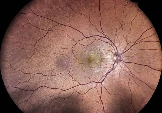
Hypertensive choroidopathy is a distinct manifestation of severe, acute blood pressure elevation affecting the choroidal circulation rather than the retinal vessels, and it is mechanistically and clinically separate from the more familiar hypertensive retinopathy.
The choroid, unlike the retina, has no autoregulatory capacity to protect itself against sudden, severe pressure surges, which is exactly why acute, malignant-range hypertension can damage choroidal vessels so abruptly and so distinctively.
This distinctive fundus appearance is genuinely useful clinically, since it can serve as an objective, visible marker of just how acutely severe an episode of hypertension has actually been, independent of whatever blood pressure number happens to be recorded at the moment of examination.
Why the Choroid Responds Differently From the Retina
Retinal vessels possess autoregulation — the ability to constrict or dilate in response to changing blood pressure to keep retinal blood flow stable — which is why hypertensive retinopathy tends to develop more gradually, through the chronic vascular changes (arteriolar narrowing, arteriovenous nicking, and eventually hemorrhages and exudates) that are the focus of this site’s separate coverage of that condition.
Choroidal vessels lack this autoregulatory mechanism, meaning a sudden, severe pressure surge is transmitted unmodified into the choroidal capillary bed, producing acute fibrinoid necrosis of the choroidal vessel walls and localized choroidal ischemia — an acute injury pattern rather than the slow, chronic remodeling seen in hypertensive retinopathy.
Clinical Settings
- Malignant hypertension — severely elevated blood pressure, often with other end-organ involvement (renal, cardiac, neurologic), representing a true hypertensive emergency
- Preeclampsia and eclampsia — pregnancy-related hypertensive disease is a particularly important context, since hypertensive choroidopathy findings in a pregnant patient can be an early ocular clue to worsening preeclampsia, and communication with the obstetric team about such findings is clinically significant
- Pheochromocytoma and other causes of acute, severe catecholamine-driven blood pressure surges
- Any other cause of acute, severe hypertensive crisis
Fundus Explorer Pro
Photograph the retinal findings described here with the phone already in your pocket — 22 D optics and built-in illumination in one handheld unit.
From Choroida — the team behind this siteClinical Findings
- Elschnig spots — small, discrete, yellow-white lesions at the level of the retinal pigment epithelium, representing focal areas of choroidal infarction, which characteristically evolve into small hyperpigmented spots with a hypopigmented halo as they heal over subsequent weeks
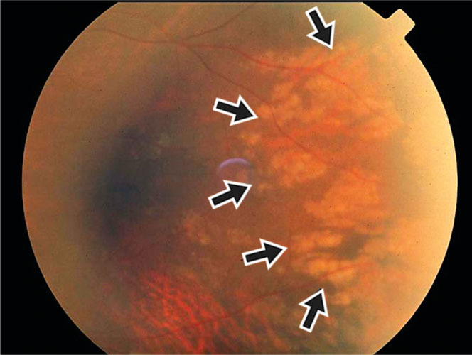
- Siegrist streaks — linear, pigmented streaks following the course of choroidal vessels, representing fibrinoid necrosis and subsequent pigmentary change along the affected vessel
- Serous retinal detachment — from disruption of the retinal pigment epithelium’s normal barrier function secondary to underlying choroidal ischemia, occurring particularly in more severe, acute cases and sometimes producing a genuinely significant, if usually reversible, degree of vision loss
- Optic disc edema, when malignant hypertension is severe enough to also produce the neuro-ophthalmic component of hypertensive emergency
- Coexisting hypertensive retinopathy findings, since severe acute hypertension frequently produces both retinal and choroidal manifestations simultaneously rather than one in isolation
Differential Diagnosis
- Central serous chorioretinopathy — also produces serous retinal detachment, but from an entirely different mechanism and typically without the severe hypertension, Elschnig spots, or Siegrist streaks that characterize the hypertensive form, discussed in its own dedicated article on this site
- Vogt-Koyanagi-Harada disease — can also produce serous retinal detachment from choroidal involvement, but as part of a distinct autoimmune, granulomatous inflammatory syndrome with its own characteristic associated findings, discussed in its own dedicated article
- Purtscher retinopathy and other acute retinal vascular insults — generally distinguished by their own specific inciting context (trauma, acute pancreatitis) and predominantly retinal, rather than choroidal, findings, discussed in this site’s separate coverage of Purtscher retinopathy
Diagnostic Evaluation
The diagnosis is largely clinical, based on recognizing the characteristic fundus findings in the setting of confirmed severe, acute hypertension.
OCT is useful for confirming and characterizing any associated serous retinal detachment or subtle RPE changes not fully appreciated on fundus exam alone, and provides a useful objective baseline for tracking resolution as treatment proceeds.
Because hypertensive choroidopathy signals a genuine hypertensive emergency, its recognition should prompt immediate coordination with internal medicine, emergency medicine, or, in pregnancy, obstetrics, for urgent blood pressure control and evaluation of other end-organ involvement.
The eye findings here are best understood as one manifestation of a systemic emergency rather than a primarily ophthalmic problem to be managed in isolation, and identifying them should trigger the same urgency as any other sign of a genuine hypertensive crisis rather than being treated as an isolated eye complaint.
Management
There is no specific ocular treatment for hypertensive choroidopathy; the eye is not treated as an isolated organ here.
Management is entirely about controlled, appropriately paced reduction of the underlying severe blood pressure elevation, coordinated with the managing medical or obstetric team, because overly rapid blood pressure reduction itself carries risks (including to cerebral and renal perfusion) that need to be balanced against the urgency of treating the hypertensive emergency.
As blood pressure is brought under control, choroidal findings — Elschnig spots evolving to their characteristic healed pigmentary pattern, and resolution of any serous retinal detachment — generally improve over subsequent weeks, and visual prognosis is usually favorable once the underlying hypertensive emergency has been adequately treated, when treatment is not significantly delayed.
Residual pigmentary change at sites of healed Elschnig spots can persist as a permanent, largely cosmetic finding even after visual function has otherwise recovered, and serves afterward as a quiet, lasting marker of the acute episode the patient experienced.


Document what you see
Two smartphone imaging tools built for everyday clinic use — one for the slit lamp, one for the fundus.
From Choroida — the team behind this siteReferences
- Bourke K, Patel MR, Prisant LM, Marcus AJ. Hypertensive choroidopathy. Journal of Clinical Hypertension.
- Hayreh SS. Hypertensive fundus changes. In: Systemic Arterial Hypertension and the Eye.
- American Academy of Ophthalmology. Basic and Clinical Science Course, Section 12: Retina and Vitreous.
Test yourself
A few questions straight from this article.
-
Hypertensive choroidopathy arises in which blood-pressure setting?
Hypertensive choroidopathy is a manifestation of severe, acute blood pressure elevation, and the fundus appearance serves as a visible marker of how acutely severe the episode has been. -
Why is the choroid particularly vulnerable to an acute hypertensive surge?
Unlike retinal vessels, choroidal vessels have no autoregulatory capacity, so a sudden severe pressure surge is transmitted straight into the choroidal capillary bed, causing fibrinoid necrosis and localized choroidal ischaemia. -
Which lesion of hypertensive choroidopathy represents focal choroidal infarction at the level of the RPE?
Elschnig spots are small discrete yellow-white lesions at the retinal pigment epithelium marking focal choroidal infarction, and they heal into hyperpigmented spots with a hypopigmented halo over weeks. -
Linear pigmented streaks following the course of choroidal vessels in hypertensive choroidopathy are called:
Siegrist streaks are linear pigmented streaks running along affected choroidal vessels, reflecting fibrinoid necrosis and the pigmentary change that follows it. -
What produces the serous retinal detachment seen in hypertensive choroidopathy?
Underlying choroidal ischaemia disrupts the normal barrier function of the retinal pigment epithelium, allowing a serous detachment that can cause significant but usually reversible vision loss. -
In a pregnant patient, hypertensive choroidopathy can be an early ocular clue to which condition?
Pregnancy-related hypertensive disease is an important context, and these choroidal findings can signal worsening preeclampsia, which makes communication with the obstetric team clinically significant. -
Which mimic of hypertensive choroidopathy causes serous detachment as part of an autoimmune granulomatous syndrome?
Vogt-Koyanagi-Harada disease also produces serous retinal detachment through choroidal involvement, but it does so as a distinct autoimmune granulomatous inflammatory syndrome with its own associated findings. -
Which investigation best confirms and tracks the serous detachment of hypertensive choroidopathy?
OCT confirms and characterizes the serous detachment and subtle RPE changes missed on fundus examination, and gives an objective baseline for following resolution during treatment. -
What forms the central management of hypertensive choroidopathy?
There is no specific ocular treatment; management is controlled reduction of the blood pressure with the medical or obstetric team, since overly rapid reduction itself threatens cerebral and renal perfusion. -
What is the usual visual outcome of hypertensive choroidopathy once the crisis is adequately treated?
As blood pressure comes under control the choroidal findings and any serous detachment improve over subsequent weeks, although residual pigmentary change at healed Elschnig spots can persist permanently.

Writes and reviews the clinical articles on Educate. Every post is checked against peer-reviewed sources before it goes live.


