Macular edema causes are many, and the finding of fluid in the macula is a sign, not a diagnosis.
The same OCT picture can also come from diabetes, vein occlusion, uveitis, cataract surgery, traction, or a medication.
A systematic approach to the cause saves the patient from treating the wrong disease.
How Macular Edema Develops
Fluid accumulates in the macula when the balance between leakage and removal is disturbed.
Also, the inner blood-retinal barrier is formed by tight junctions between retinal capillary endothelial cells, and the outer barrier by the retinal pigment epithelium.
Damage to either barrier, or dysfunction of the Müller cells and the pigment epithelium that remove fluid, leads to accumulation.
Fluid collects in the outer plexiform and inner nuclear layers, which form cystoid spaces, or under the retina as a serous detachment.
Chronic edema damages photoreceptors and the foveal architecture, so delay in finding the cause can cost vision.
Macular Edema Causes by Category
- Retinal vascular disease: diabetic retinopathy, retinal vein occlusion, hypertensive retinopathy, macular telangiectasia, Coats disease, retinal macroaneurysm, and radiation retinopathy (see diabetic macular edema and central retinal vein occlusion)
- Inflammation: anterior, intermediate, and posterior uveitis, birdshot chorioretinopathy, and sarcoidosis (see uveitic macular edema)
- After surgery: pseudophakic cystoid macular edema after cataract surgery, and after vitrectomy or retinal detachment surgery (see Irvine-Gass syndrome)
- Traction: epiretinal membrane and vitreomacular traction (see epiretinal membrane and vitreomacular traction)
- Choroidal and degenerative disease: neovascular AMD, polypoidal choroidal vasculopathy, and myopic choroidal neovascularization
- Hereditary conditions: retinitis pigmentosa, and some dystrophies
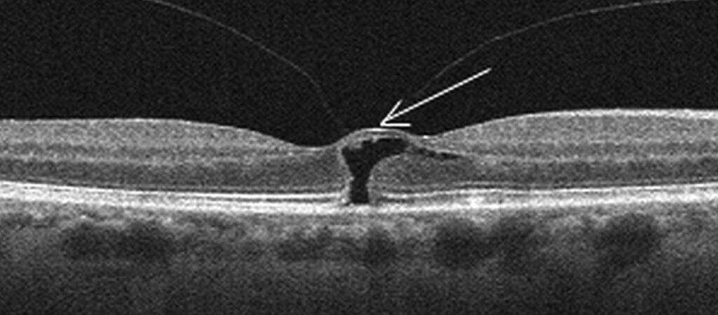
- Optic nerve head anomalies: optic disc pit maculopathy (see optic pit maculopathy)
- Medications: prostaglandin analogs, nicotinic acid, taxanes, glitazones, and fingolimod
Fundus Explorer Pro
Photograph the retinal findings described here with the phone already in your pocket — 22 D optics and built-in illumination in one handheld unit.
From Choroida — the team behind this sitePatterns That Point to the Cause
OCT shows diffuse thickening with cystoid spaces in vascular disease and uveitis, with a typical petalloid pattern on fluorescein angiography.
Tractional edema has a visible membrane or adhesion, often with a distorted foveal contour and an elevated surface.
Serous detachment under the fovea is more common in vein occlusion and in choroidal disease.
Cystic spaces without leakage on angiography suggest a nonleaking cause such as niacin maculopathy, taxane toxicity, or X-linked retinoschisis (see X-linked retinoschisis).
Edema with hemorrhages, exudates, or vessel changes points to vascular disease, while edema with vitreous cells and keratic precipitates suggests inflammation.
Evaluation for Macular Edema Causes
Take a history of diabetes, hypertension, eye surgery, systemic inflammatory disease, and medications.
Examine the anterior segment and the vitreous for inflammation, the lens status, and the fundus for hemorrhages, exudates, and membranes.
Use OCT to define the pattern, and fluorescein angiography to show the source of leakage when the cause is not clear (see fluorescein angiography patterns).
OCT angiography can show nonperfusion and neovascular membranes without dye (see OCT angiography).
Order blood tests for glucose and inflammatory disease according to the picture.
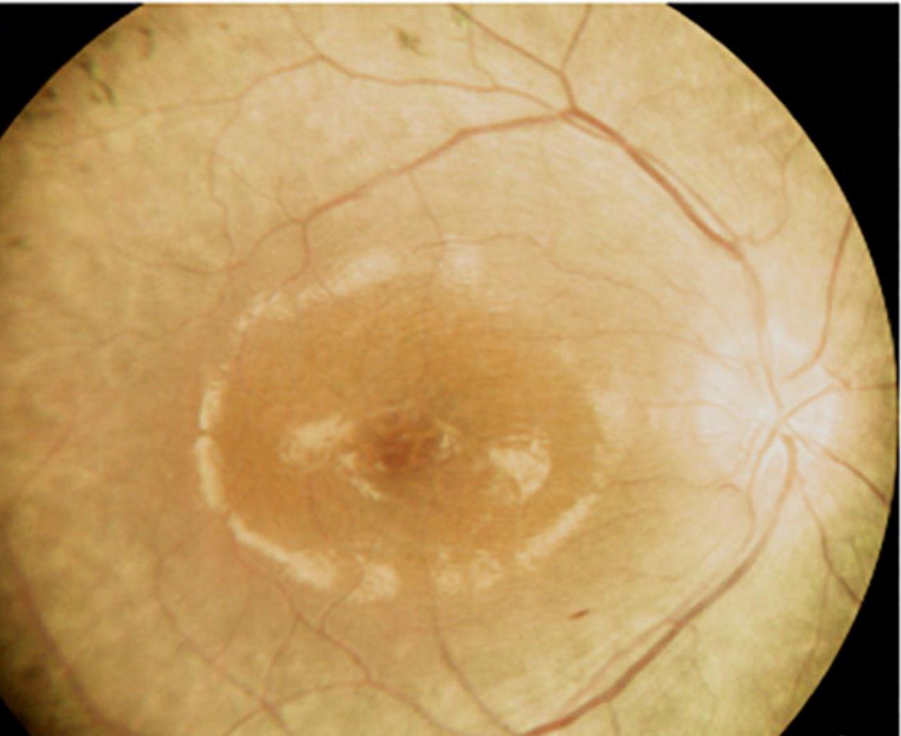
Macular Edema Causes: Management Principles
Treat the cause first.
In diabetic and vein occlusion edema, anti-VEGF drugs are first-line, with steroids and laser as alternatives (see anti-VEGF therapy).
Inflammatory edema needs control of the uveitis, with topical, periocular, or systemic corticosteroids and steroid-sparing agents.
Pseudophakic edema responds to topical nonsteroidal and steroid drops, with other options for persistent cases.
Tractional edema may require surgery to remove the membrane or relieve traction.
Drug-induced edema often improves when the drug is stopped, in consultation with the prescribing physician.
Carbonic anhydrase inhibitors can help in retinitis pigmentosa and some other hereditary conditions.
Counselling
Explain that edema is a sign of an underlying problem, that finding the cause guides treatment, and that vision often improves when the cause is treated.
Emphasize regular follow-up with OCT, because recurrence is common.
Common Pitfalls
- Treating every cystoid change as diabetic macular edema without asking about other causes
- Missing a tractional component, such as a subtle epiretinal membrane or vitreomacular adhesion, that explains a poor response to injections
- Forgetting medications, including niacin, taxanes, glitazones, and prostaglandin analogs in aphakic or pseudophakic eyes
- Overlooking uveitis in a patient with unexplained edema, particularly in young patients and in those with mild inflammation
- Failing to look for a hereditary cause when edema is bilateral and has no leakage on angiography
Monitoring Response
OCT is the main tool for judging response, with central subfield thickness and the presence of intraretinal fluid as outcome measures.
Acuity does not always follow thickness, because chronic edema damages photoreceptors and the foveal contour.
Look at the outer retina and the ellipsoid zone as well as the thickness, since they predict the potential for recovery (see ellipsoid zone disruption).
If edema persists despite treatment, reconsider the diagnosis, look for a second cause, and consider changing the drug or adding another therapy.
For the latest studies, see the PubMed results for macular edema causes.


Document what you see
Two smartphone imaging tools built for everyday clinic use — one for the slit lamp, one for the fundus.
From Choroida — the team behind this siteReferences
- Rotsos TG, Moschos MM. Cystoid macular edema. Clin Ophthalmol. 2008;2:919-930.
- Yanoff M, Duker JS, eds. Ophthalmology. 5th ed. Elsevier; 2019.
- Ryan SJ, Schachat AP, Wilkinson CP, et al, eds. Retina. 6th ed. Elsevier; 2018.
- American Academy of Ophthalmology. Basic and Clinical Science Course, Section 12: Retina and Vitreous. AAO; current edition.

Writes and reviews the clinical articles on Educate. Every post is checked against peer-reviewed sources before it goes live.