Aqueous-deficient dry eye is the form of dry eye disease in which the lacrimal glands fail to produce enough of the watery component of the tear film.
It is less common than evaporative dry eye, but it tends to be more severe.
It often points to a systemic disease that needs its own work-up.
Separating it from evaporative disease changes the tests, the treatment, and sometimes the referral.
What Is Aqueous-Deficient Dry Eye?
The tear film has a lipid layer from the meibomian glands, a watery layer from the lacrimal glands, and a mucin layer from the conjunctival goblet cells and the epithelium.
In aqueous-deficient disease, the watery layer is also reduced, the tear volume falls, and the osmolarity of the tears rises.
The raised osmolarity damages the ocular surface and triggers inflammation, which in turn damages the lacrimal gland and the surface further.
In addition, the Tear Film and Ocular Surface Society report on dry eye divides the aqueous-deficient group into Sjögren syndrome dry eye and non-Sjögren dry eye.
Many patients have a mixed picture, with some degree of meibomian gland dysfunction as well.
The aqueous deficiency may not be the only problem.
Causes of Aqueous-Deficient Dry Eye
- Sjögren syndrome: autoimmune infiltration of the lacrimal and salivary glands (see Sjögren syndrome)
- Age-related lacrimal gland dysfunction: fibrosis and obstruction of the ducts
- Lacrimal gland infiltration: sarcoidosis, lymphoma, and amyloid
- Ocular graft-versus-host disease and cicatrizing disease: destruction of the ducts (see ocular graft-versus-host disease and ocular cicatricial pemphigoid)
- Reduced reflex tearing: contact lens wear, refractive surgery, and neurotrophic corneas
- Drugs: antihistamines, anticholinergics, antidepressants, diuretics, and some isotretinoin regimens
- Radiation, surgery, and trauma to the lacrimal glands
All-fit Slit-Lamp Adapter
Record and share exactly what you see at the slit lamp. One adapter fits any slit lamp or surgical microscope — and any smartphone.
From Choroida — the team behind this siteClinical Features of Aqueous-Deficient Dry Eye
Patients complain of dryness, burning, grittiness, and a foreign body sensation that worsens through the day and in dry or windy places.
Paradoxically, some complain of watering because the cornea becomes irritated and triggers reflex tearing.
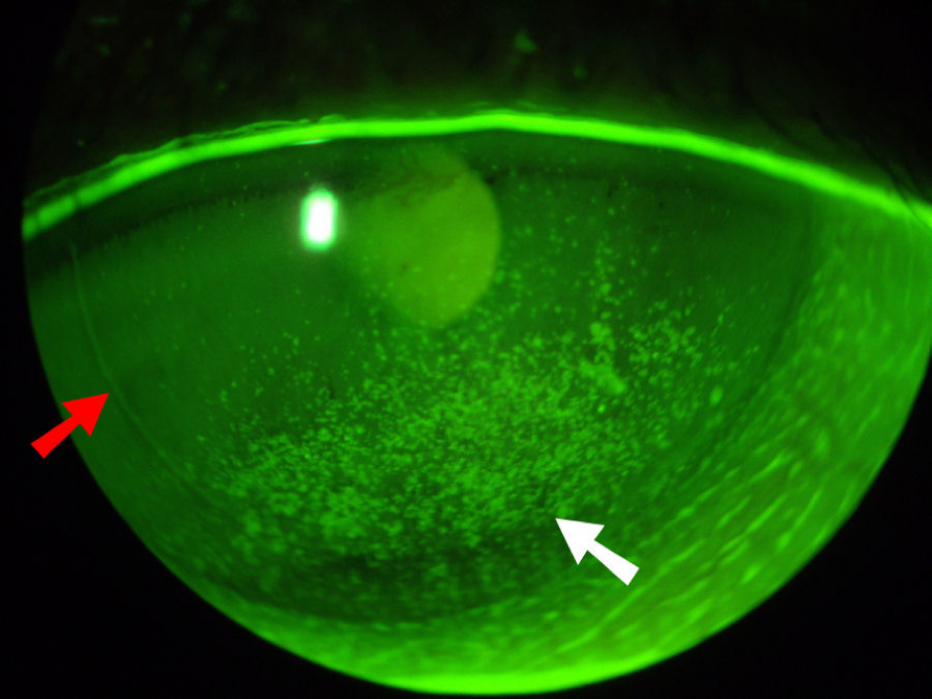
On examination, the tear meniscus is low or absent, and debris and mucus strands may also be present in the tear film.
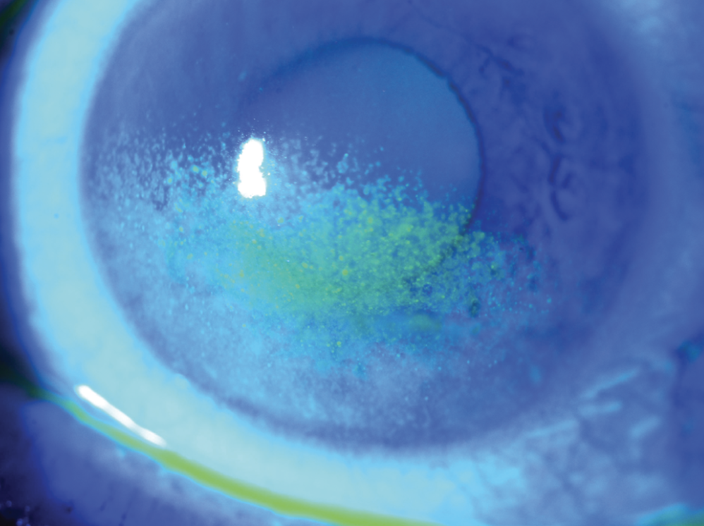
Also, fluorescein staining shows punctate keratopathy in the interpalpebral zone, and lissamine green stains the conjunctiva in a band pattern.
Filaments may form in severe disease (see filamentary keratitis).
Investigations
- Schirmer test without anesthesia, with low values pointing to aqueous deficiency
- Tear meniscus height measured with the slit lamp or OCT
- Tear osmolarity, when the equipment is available
- Fluorescein and lissamine green staining, graded on a standard scale
- Tear break-up time to look for coexisting evaporative disease
- Serology for anti-Ro and anti-La antibodies, antinuclear antibodies, and rheumatoid factor in suspected Sjögren syndrome
A patient with severe aqueous deficiency and no clear cause deserves a systemic work-up, because dry eye can be the first sign of Sjögren syndrome or sarcoidosis.
Management
Treatment follows a stepwise approach that increases in intensity with severity (see dry eye disease).
Also, begin with education, environmental changes, and preservative-free artificial tears, and treat any lid disease.
Add punctal occlusion to retain the tears the patient still produces (see punctal plugs).
Anti-inflammatory therapy, such as topical cyclosporine, lifitegrast, or a short course of a low-dose corticosteroid, addresses the inflammatory loop.
In Sjögren syndrome, oral secretagogues such as pilocarpine or cevimeline can increase secretion if the patient tolerates the side effects.
Autologous serum drops, and scleral contact lenses that hold a reservoir of fluid over the cornea, are valuable for severe disease (see scleral contact lens fitting).
Surgical options include permanent punctal occlusion and tarsorrhaphy for the most severe cases.
Counselling
Explain that the condition is chronic, that treatment controls it but does not cure it, and that symptoms may fluctuate with the environment.
Patients with Sjögren syndrome need regular review by a rheumatologist and awareness of the increased risk of lymphoma, which is small but real.
Advise them to report any new redness with pain or a drop in vision, which could indicate an infected corneal ulcer, a risk in these dry eyes.
Practical Pearls
A patient with a very low Schirmer value and dry mouth should be asked about joint pain, dental problems, and fatigue, because Sjögren syndrome is often diagnosed late.
Review the medication list at every visit, since stopping or switching an anticholinergic drug sometimes improves tear production.
Remember that severe aqueous deficiency raises the risk of corneal infection and melting, so a painful red eye in such a patient needs a prompt slit-lamp examination.
Keep a simple record of the symptom score, staining grade, and Schirmer value at each visit, because the combination gives a clearer picture of progress than any single number.
For the latest studies, see the PubMed results for aqueous-deficient dry eye.


Document what you see
Two smartphone imaging tools built for everyday clinic use — one for the slit lamp, one for the fundus.
From Choroida — the team behind this siteReferences
- Craig JP, Nichols KK, Akpek EK, et al. TFOS DEWS II definition and classification report. Ocul Surf. 2017;15:276-283.
- Bron AJ, de Paiva CS, Chauhan SK, et al. TFOS DEWS II pathophysiology report. Ocul Surf. 2017;15:438-510.
- Jones L, Downie LE, Korb D, et al. TFOS DEWS II management and therapy report. Ocul Surf. 2017;15:575-628.
- American Academy of Ophthalmology. Basic and Clinical Science Course, Section 8: External Disease and Cornea. AAO; current edition.

Writes and reviews the clinical articles on Educate. Every post is checked against peer-reviewed sources before it goes live.