
Diabetic macular edema (DME) is fluid accumulation in the macula caused by breakdown of the blood-retinal barrier in a diabetic eye.
It is the most common cause of central vision loss in people with diabetes, and it can develop at any stage of diabetic retinopathy.
Treatment has changed a great deal over the last fifteen years, and the decision now rests on visual acuity, OCT anatomy, and the response to the first agent, with the appearance of leakage counting for less.
This article covers how DME develops, how it is graded, and how to choose between observation, anti-VEGF injections, corticosteroids, and laser.
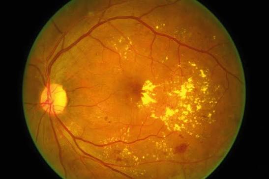
What Is Diabetic Macular Edema?
DME is retinal thickening from intraretinal and sometimes subretinal fluid that accumulates in the macular region.
The older ETDRS term was clinically significant macular edema (CSME), defined on stereoscopic biomicroscopy or photographs by any of the following:
- Retinal thickening at or within 500 µm of the foveal center
- Hard exudates at or within 500 µm of the center, with adjacent retinal thickening
- A zone of retinal thickening one disc area or larger, any part of which lies within one disc diameter of the center
Current practice uses OCT instead.
DME is called center-involving (CI-DME) when the central subfield is thickened above the normal range for the device, and non-center-involving when the fluid spares the central 1 mm.
The distinction matters because almost all the modern trial evidence applies to center-involving disease.
Pathogenesis
Chronic hyperglycemia damages the retinal microvasculature through pericyte loss, endothelial dysfunction, leukostasis, and low-grade inflammation.
VEGF, IL-6, and other cytokines increase vascular permeability, and the tight junctions of the inner blood-retinal barrier loosen.
Fluid leaks faster than the RPE pump and Müller cells can remove it, so it collects in the outer plexiform and inner nuclear layers first.
In some eyes the fluid also enters the subretinal space.
Vitreomacular traction and an epiretinal membrane can add a mechanical component, and systemic hypertension, dyslipidemia, renal disease, and fluid retention all worsen the picture.
Fundus Explorer Pro
Photograph the retinal findings described here with the phone already in your pocket — 22 D optics and built-in illumination in one handheld unit.
From Choroida — the team behind this siteClinical Presentation
Early DME is often asymptomatic, especially when the fovea is spared.
Central involvement produces blurred vision, difficulty reading, reduced contrast, and mild metamorphopsia.
On examination the retina looks thickened and dull, with microaneurysms, dot-blot hemorrhages, and hard exudates.
A circinate ring of hard exudates around a cluster of leaking microaneurysms marks a focal source of leakage.
Visual acuity may be normal in eyes with good anatomy, which is why OCT screening is worth doing in every diabetic eye with retinopathy.
Imaging
OCT
OCT is the main tool for diagnosis and follow-up.
It shows cystoid spaces, diffuse thickening, subretinal fluid, hyperreflective foci, and any vitreomacular interface abnormality.
Several biomarkers help predict visual outcome:
- Disorganization of the retinal inner layers (DRIL), which correlates with poor acuity even after the edema resolves
- Ellipsoid zone and external limiting membrane disruption
- Central subfield thickness, which correlates only loosely with acuity
- Hyperreflective foci, which may reflect lipid extravasation or inflammation
Fluorescein Angiography
Fluorescein angiography is not needed to diagnose DME, but it identifies leaking microaneurysms before focal laser and shows macular ischemia, which limits visual potential (see diabetic macular ischemia).
OCT Angiography
OCT angiography shows enlargement and irregularity of the foveal avascular zone and capillary dropout without dye, which helps in patients who cannot have fluorescein (see OCT angiography).
Differential Diagnosis
Not every thickened macula in a diabetic patient is DME.
Other causes of macular edema can coexist or mimic it:
- Retinal vein occlusion
- Pseudophakic cystoid macular edema
- Uveitic macular edema
- Hypertensive retinopathy and radiation retinopathy
- Vitreomacular traction or epiretinal membrane
- Drug-induced edema, such as from niacin or taxanes
- Neovascular AMD with intraretinal fluid
Asymmetric edema, edema out of proportion to the retinopathy, or poor response to anti-VEGF should prompt a second look for one of these.
Management
Systemic Control
Glycemic, blood pressure, and lipid control reduce the risk of onset and progression.
Very rapid lowering of HbA1c can transiently worsen retinopathy, so patients with advanced disease need eye examination during intensive control.
In ACCORD Eye, adding fenofibrate to simvastatin reduced the rate of retinopathy progression in type 2 diabetes.
Observation
DRCR Protocol V randomized eyes with center-involving DME and good acuity (20/25 or better) to aflibercept, laser, or observation with aflibercept rescue.
Visual acuity at two years was similar in all three groups, and only a minority of the observed eyes needed rescue treatment.
Careful observation is therefore reasonable when vision is good and follow-up is dependable.
Anti-VEGF Therapy
Anti-VEGF agents are first-line for center-involving DME with visual impairment.
In DRCR Protocol T, aflibercept, bevacizumab, and ranibizumab all improved acuity.
Aflibercept gave larger gains than the other two when baseline acuity was 20/50 or worse at one year, and the difference between aflibercept and ranibizumab was smaller at two years.
Faricimab, a bispecific antibody that blocks VEGF-A and angiopoietin-2, achieved noninferior acuity gains to aflibercept in the YOSEMITE and RHINE trials, and a proportion of patients could be extended to 16-week dosing.
Monthly loading for the first several injections, followed by treat-and-extend or PRN dosing, is the usual approach (see anti-VEGF therapy and intravitreal injection therapy).
Anti-VEGF therapy carries the small risks of endophthalmitis, intraocular inflammation, and a transient IOP rise.
It requires many visits over several years, and patients who stop early tend to lose the gains.
Corticosteroids
Corticosteroids suppress both VEGF and inflammatory mediators, so they help eyes with a partial anti-VEGF response.
The dexamethasone implant improved acuity in the MEAD trial, with about 22% of treated eyes gaining 15 or more letters compared with 12% of sham eyes, but cataract developed in most phakic eyes.
IOP elevation needs monitoring and sometimes treatment.
Steroids fit best in pseudophakic eyes, in patients who cannot attend frequent visits, and in eyes with a suboptimal anti-VEGF response (see steroids in resistant DME).
Laser Photocoagulation
The ETDRS showed that focal or grid laser roughly halved the three-year rate of moderate visual loss, from about 24% to 12%.
Laser stabilizes vision but rarely improves it, so it is now used as an adjunct for focal leakage from microaneurysms outside the fovea and is seldom the primary treatment.
Subthreshold micropulse laser is used in some centers, although the evidence for it is weaker.
Vitrectomy
Vitrectomy with membrane peeling is considered when there is vitreomacular traction, a taut epiretinal membrane, or persistent edema that has failed medical therapy and has a mechanical component on OCT.
Prognosis
Most eyes treated promptly with anti-VEGF keep or improve their vision.
Baseline acuity, DRIL, ellipsoid zone integrity, macular ischemia, and treatment adherence predict the final result.
Eyes with long-standing edema and outer retinal damage recover less, and vision may plateau even when the retina looks dry.
Patients need to understand that the aim is to preserve vision and that treatment is often long-term.


Document what you see
Two smartphone imaging tools built for everyday clinic use — one for the slit lamp, one for the fundus.
From Choroida — the team behind this siteReferences
- Early Treatment Diabetic Retinopathy Study Research Group. Photocoagulation for diabetic macular edema. ETDRS report number 1. Arch Ophthalmol. 1985;103:1796-1806.
- Diabetic Retinopathy Clinical Research Network. Aflibercept, bevacizumab, or ranibizumab for diabetic macular edema. N Engl J Med. 2015;372:1193-1203.
- Baker CW, Glassman AR, Beaulieu WT, et al. Effect of initial management with aflibercept vs laser photocoagulation vs observation on vision loss among patients with diabetic macular edema involving the center of the macula and good visual acuity: a randomized clinical trial. JAMA. 2019;321:1880-1894.
- Boyer DS, Yoon YH, Belfort R Jr, et al. Three-year, randomized, sham-controlled trial of dexamethasone intravitreal implant in patients with diabetic macular edema. Ophthalmology. 2014;121:1904-1914.
- Wykoff CC, Abreu F, Adamis AP, et al. Efficacy, durability, and safety of intravitreal faricimab with extended dosing up to every 16 weeks in patients with diabetic macular oedema (YOSEMITE and RHINE): two randomised, double-masked, phase 3 trials. Lancet. 2022;399:741-755.
- ACCORD Study Group and ACCORD Eye Study Group. Effects of medical therapies on retinopathy progression in type 2 diabetes. N Engl J Med. 2010;363:233-244.
- Duh EJ, Sun JK, Stitt AW. Diabetic retinopathy: current understanding, mechanisms, and treatment strategies. JCI Insight. 2017;2(14):e93751.
Test yourself
A few questions straight from this article.
-
In diabetic macular edema, what defines center-involving disease?
DME is center-involving when the central subfield is thickened above the normal range for the OCT device, and non-center-involving when fluid spares the central 1 mm. Almost all modern trial evidence applies to center-involving disease. -
Where does fluid first collect within the retina in diabetic macular edema?
Fluid leaks faster than the RPE pump and Müller cells can clear it, so it collects first in the outer plexiform and inner nuclear layers; in some eyes it later enters the subretinal space. -
What does a circinate ring of hard exudates in diabetic macular edema indicate?
A circinate ring of hard exudates surrounds a cluster of leaking microaneurysms and marks a focal source of leakage. -
Which OCT biomarker predicts poor acuity in diabetic macular edema even after the edema resolves?
DRIL correlates with poor acuity even after the edema has resolved, whereas central subfield thickness correlates only loosely with visual acuity. -
Which feature should prompt a search for a cause of macular edema other than diabetes?
Asymmetric edema, edema out of proportion to the retinopathy, or a poor anti-VEGF response should prompt a second look for vein occlusion, uveitis, traction or another cause. -
DRCR Protocol V studied center-involving DME with acuity of 20/25 or better. What did it show at two years?
Protocol V randomized these eyes to aflibercept, laser, or observation with aflibercept rescue. Acuity at two years was similar in all three groups and only a minority of observed eyes needed rescue, so careful observation is reasonable when vision is good and follow-up is dependable. -
In DRCR Protocol T, which agent gave larger one-year acuity gains when baseline acuity was 20/50 or worse?
All three agents studied improved acuity, but aflibercept gave larger gains than bevacizumab and ranibizumab at one year when baseline acuity was 20/50 or worse; the difference from ranibizumab was smaller at two years. -
Which eyes with diabetic macular edema are best suited to a dexamethasone implant?
Steroids suit pseudophakic eyes, patients who cannot attend frequent visits, and eyes with a suboptimal anti-VEGF response. Cataract developed in most phakic eyes in the MEAD trial, and IOP elevation needs monitoring. -
What effect did focal or grid laser have on moderate visual loss in the ETDRS?
The ETDRS showed focal or grid laser roughly halved the three-year rate of moderate visual loss, from about 24% to 12%. Laser stabilizes vision but rarely improves it, so it is now an adjunct rather than primary treatment. -
Why may vision plateau in diabetic macular edema even when the retina looks dry?
Eyes with long-standing edema and outer retinal damage recover less, and vision may plateau even when the retina appears dry. Baseline acuity, DRIL, ellipsoid zone integrity, ischemia and adherence all predict the final result.

Writes and reviews the clinical articles on Educate. Every post is checked against peer-reviewed sources before it goes live.


