Not every cataract is a matter of aging.
A complicated cataract is one that develops because of another disease within the eye, and recognizing it matters because the underlying condition shapes the surgical plan, the expected outcome, and the advice given to the patient.
The typical appearance
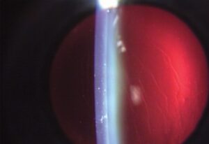
The lens changes begin in the posterior cortex, near the posterior pole.
At first there are fine, scattered granules and a polychromatic or “rainbow” luster visible on retroillumination.
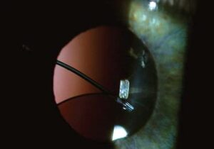
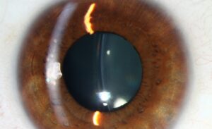
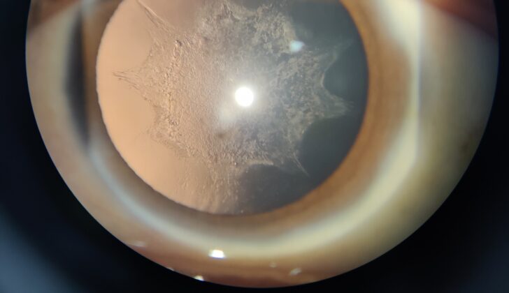
As the opacity grows, it forms a posterior subcapsular plaque that may extend towards the equator, and later the entire lens may become opaque.
In some cases, a “bread-crumb” appearance is described.
Patients complain of glare and decreased vision in bright light, and symptoms are often out of proportion to the apparent size of the opacity because it lies close to the nodal point of the eye (see posterior subcapsular cataract).
Common underlying causes
- Chronic uveitis, including juvenile idiopathic arthritis-associated uveitis, Fuchs heterochromic iridocyclitis, and HLA-B27 disease, in which inflammation and steroid treatment both contribute (see Fuchs heterochromic iridocyclitis)
- Retinitis pigmentosa, in which posterior subcapsular cataract appears in up to half of patients (see retinitis pigmentosa management)
- High myopia, which promotes early nuclear and posterior subcapsular changes
- Chronic or acute angle-closure glaucoma, which can cause glaukomflecken, small anterior subcapsular opacities, after an acute attack
- Intraocular tumors, long-standing retinal detachment, and hypotony
- Ocular ischemia and prior intraocular surgery, including vitrectomy
- Penetrating or blunt trauma
- Exposure to toxic agents such as chlorpromazine or amiodarone
All-fit Slit-Lamp Adapter
Record and share exactly what you see at the slit lamp. One adapter fits any slit lamp or surgical microscope — and any smartphone.
From Choroida — the team behind this siteHow it differs from age-related cataract
Age-related cataract tends to affect the nucleus and cortex first, and it is bilateral and symmetric.
Complicated cataract starts in the posterior subcapsular area, and it is often unilateral or asymmetric, with an onset that is early for the patient’s age.
The pattern gives a clue that a search for another disease is needed.
What to look for
- Anterior chamber cells and flare, posterior synechiae, and keratic precipitates, indicating uveitis
- Pigmentary changes, vessel attenuation and bone-spicule pigmentation in the peripheral retina
- Axial length and optic disc appearance in myopia and glaucoma
- Heterochromia, stellate keratic precipitates and absent synechiae in Fuchs syndrome
When the fundus cannot be seen, B-scan ultrasonography is used to exclude a tumor or detachment.
Surgical considerations
Operating on a complicated cataract demands preparation.
- Control inflammation for at least three months before surgery in uveitis, and use perioperative corticosteroid cover
- Expect posterior synechiae, small pupils, and fibrotic capsules, and have pupil expansion devices ready
- In retinitis pigmentosa, zonular weakness and a tendency to capsule phimosis may need capsular tension rings
- Inform the patient that the visual result may be limited by the underlying disease, for example macular edema in uveitis or retinal degeneration
- Plan for IOL choice carefully, since some IOLs are better tolerated in uveitic eyes
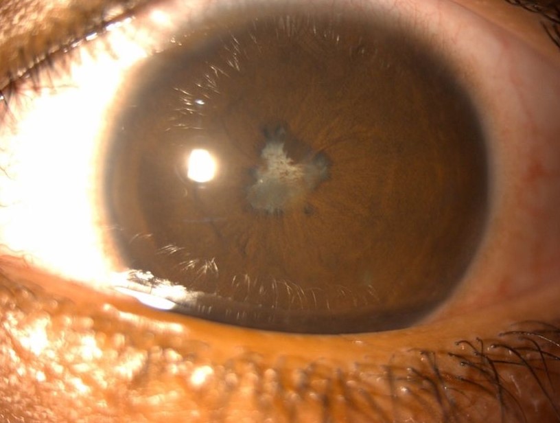
Finding the underlying cause
A systematic search is needed when a younger patient presents with posterior subcapsular changes in one eye. Ask about eye inflammation, injuries, surgery, family history of retinal dystrophy, long-term drug treatment, and metabolic disease. Examine the iris for atrophy, synechiae, and heterochromia, and examine the angle and the pressure. Dilate fully and examine the retina, using ultrasound if the view is poor.
Planning the operation
Complicated cataracts can have unexpected features. In uveitic eyes, expect posterior synechiae, a fibrotic capsule, and a risk of postoperative inflammation and macular edema. In retinitis pigmentosa, the zonules may be weak, and the capsulorhexis should be generous, with a capsular tension ring in reserve. In high myopia, the anterior chamber may be deep and the posterior capsule may be fragile, and the retina needs a careful examination before and after surgery.
Setting expectations is important, since the best-corrected vision after surgery may be limited by the disease that caused the cataract. Even so, many patients benefit through improved contrast, color, and quality of life.
Counselling and expectations
Explain to the patient that the cataract is a symptom of another eye condition, that surgery can improve vision but may not restore it fully, and that monitoring of the underlying disease continues after surgery. A realistic discussion before the operation avoids disappointment and builds trust, particularly in patients with uveitis or retinal degeneration.
Prognosis
Many patients achieve a good result, and surgery can improve quality of life even when the potential vision is limited.
Counselling about realistic expectations and close postoperative follow-up are central to success.


Document what you see
Two smartphone imaging tools built for everyday clinic use — one for the slit lamp, one for the fundus.
From Choroida — the team behind this siteReferences
- Foster CS, Fong LP, Singh G. Cataract surgery and intraocular lens implantation in patients with uveitis. Ophthalmology. 1989;96:281-287.
- Jackson H, Garway-Heath D, Rosen P, Bird AC, Tuft SJ. Outcome of cataract surgery in patients with retinitis pigmentosa. Br J Ophthalmol. 2001;85:936-938.
- Okhravi N, Lightman SL, Towler HM. Assessment of visual outcome after cataract surgery in patients with uveitis. Ophthalmology. 1999;106:710-722.
- Elgohary MA, McCluskey PJ, Towler HM, et al. Outcome of phacoemulsification in patients with uveitis. Br J Ophthalmol. 2007;91:916-921.
Test yourself
A few questions straight from this article.
-
What defines a complicated cataract?
A complicated cataract develops because of another disease within the eye, and that underlying condition shapes the surgical plan, the expected outcome and the advice given. -
Which retroillumination appearance is characteristic of an early complicated cataract?
The earliest change is fine scattered granules with a polychromatic or rainbow luster on retroillumination, which later forms a posterior subcapsular plaque. -
Which pattern suggests a complicated rather than an age-related cataract?
Age-related cataract affects the nucleus and cortex first and is bilateral and symmetric, whereas a complicated cataract starts posterior subcapsularly and is often unilateral or asymmetric and early for the patient's age. -
In retinitis pigmentosa, posterior subcapsular cataract appears in roughly what proportion of patients?
Posterior subcapsular cataract is reported in up to half of patients with retinitis pigmentosa. -
What are glaukomflecken, seen after an acute angle-closure attack?
Glaukomflecken are small anterior subcapsular lens opacities that follow an acute angle-closure attack. -
Which combination of signs points to Fuchs heterochromic iridocyclitis?
Fuchs syndrome is marked by heterochromia, stellate keratic precipitates and the absence of synechiae, which separates it from other causes of uveitic cataract. -
How long should inflammation be controlled before cataract surgery in a uveitic eye?
Inflammation should be controlled for at least three months before surgery in uveitis, with perioperative corticosteroid cover. -
Which device should be held in reserve for cataract surgery in retinitis pigmentosa?
In retinitis pigmentosa the zonules may be weak and the capsule tends to undergo phimosis, so the capsulorhexis should be generous with a capsular tension ring available. -
Which drugs are listed as toxic causes of a complicated cataract?
Exposure to toxic agents such as chlorpromazine or amiodarone is among the recognised causes of a complicated cataract. -
What should a patient with a complicated cataract be told about the expected outcome of surgery?
Best-corrected vision may be limited by the disease that caused the cataract, yet many patients benefit through improved contrast, colour and quality of life.

Writes and reviews the clinical articles on Educate. Every post is checked against peer-reviewed sources before it goes live.