
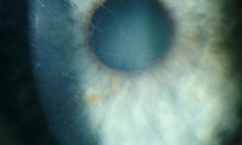
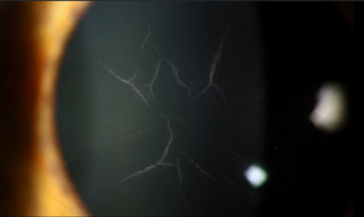
Lattice corneal dystrophy produces one of the most visually distinctive patterns in all of corneal disease, thin, glassy, branching refractile lines running through the stroma like the threads of a spider’s web, and recognizing this pattern at the slit lamp usually settles the diagnosis on sight, well before any genetic or histopathologic confirmation is needed.
What’s Actually Accumulating
Lattice corneal dystrophy results from the progressive deposition of amyloid, an abnormal, misfolded protein, within the corneal stroma, and this amyloid deposition is what produces both the characteristic refractile branching lines visible on slit-lamp examination and the progressive stromal haze that develops as the disease advances.
Most cases result from mutations in the TGFBI gene, and inheritance is typically autosomal dominant, though the specific mutation, age of onset, and rate of progression vary across the different recognized subtypes of lattice dystrophy.
Clinical Presentation
- Thin, branching, glassy refractile lines within the stroma, most easily seen with retroillumination or broad, oblique slit-lamp illumination, typically most numerous centrally and becoming sparser toward the periphery
- Onset is typically in the first or second decade of life for the classic type 1 form, with slow progression over subsequent decades
- Bilateral, though asymmetry between the two eyes is common
- Progressive stromal haze develops over time as the amyloid deposits become more extensive and confluent, eventually reducing vision independent of any acute erosion episode
- Recurrent corneal erosion is a prominent and often the most immediately troublesome feature, since the amyloid deposits disrupt normal epithelial basement membrane adhesion, producing the same painful, often nocturnal, breakdown episodes seen in primary recurrent erosion syndrome
All-fit Slit-Lamp Adapter
Record and share exactly what you see at the slit lamp. One adapter fits any slit lamp or surgical microscope — and any smartphone.
From Choroida — the team behind this siteDistinguishing Lattice From Other Stromal Dystrophies
- Granular corneal dystrophy produces discrete, breadcrumb-like white deposits with clear stroma between them, a visually distinct pattern from lattice’s branching lines
- Macular corneal dystrophy produces a more diffuse, cloudy stromal haze extending to the limbus, without the sharply refractile branching lines characteristic of lattice
- Some patients have combined granular-lattice deposits, historically termed Avellino dystrophy, now recognized as a specific TGFBI mutation producing overlapping features of both patterns
Systemic Associations
While most lattice corneal dystrophy is an isolated ocular condition, a rarer form, lattice dystrophy type 2, is associated with systemic amyloidosis (Meretoja syndrome), and recognizing systemic features such as cranial and peripheral neuropathy, lax skin, and other manifestations of systemic amyloid deposition in a patient with lattice-pattern corneal findings should prompt evaluation for this broader syndrome rather than assuming an isolated ocular dystrophy.
Diagnosis
Diagnosis is typically made clinically based on the characteristic slit-lamp appearance, particularly the branching refractile lines on retroillumination, combined with a family history consistent with autosomal dominant inheritance.
Genetic testing for TGFBI mutations can confirm the diagnosis and specific subtype, and is particularly useful for family counseling and when systemic amyloidosis is a concern.
Management
Managing Recurrent Erosion
Recurrent erosion episodes are managed with the same stepwise approach used for primary recurrent corneal erosion syndrome: lubrication, hypertonic saline, bandage contact lens for acute episodes, and, for frequent or severe recurrence, anterior stromal micropuncture or phototherapeutic keratectomy (see recurrent corneal erosion syndrome).
Managing Progressive Visual Loss
- Phototherapeutic keratectomy can also be used to ablate more superficial amyloid deposits and improve both surface irregularity and some degree of stromal clarity, though deeper deposits are not reached by this technique
- Corneal transplantation, whether lamellar or full-thickness, is considered once stromal haze has progressed enough to significantly impair vision, with deep anterior lamellar keratoplasty often preferred when the endothelium remains healthy (see deep anterior lamellar keratoplasty)
Recurrence After Transplantation
A notable feature of lattice corneal dystrophy is that amyloid deposits can recur in the donor graft tissue over subsequent years to decades, since the underlying genetic and metabolic abnormality driving amyloid production is systemic to the patient’s own corneal epithelium and keratocyte activity, not eliminated by replacing the central stromal tissue alone.
Prognosis
Lattice corneal dystrophy is a slowly progressive condition, and most patients maintain functional vision for many years, with recurrent erosion often being the more immediately troublesome symptom before visually significant stromal haze develops.
Corneal transplantation restores vision effectively when needed, though patients should be counseled about the realistic possibility of disease recurrence in the graft over the long term, supporting the value of ongoing follow-up even after an apparently successful transplant.


Document what you see
Two smartphone imaging tools built for everyday clinic use — one for the slit lamp, one for the fundus.
From Choroida — the team behind this siteReferences
- Klintworth GK. Corneal dystrophies. Orphanet J Rare Dis. 2009;4:7.
- Weiss JS, Moller HU, Aldave AJ, et al. IC3D classification of corneal dystrophies–edition 2. Cornea. 2015;34:117-159.
- Meretoja J. Familial systemic paramyloidosis with lattice dystrophy of the cornea, progressive cranial neuropathy, skin changes and various internal symptoms. Ann Clin Res. 1969;1:314-324.
- Stewart HS, Ridgway AE, Dixon MJ, Bonshek R, Parveen R, Black G. Heterogeneity in granular corneal dystrophy: identification of three causative mutations in the TGFBI (BIGH3) gene. Hum Mutat. 1999;14:126-132.
