
Sensory strabismus is eye misalignment that develops as a secondary consequence of poor vision in one eye, rather than as a primary problem of ocular alignment or extraocular muscle function.
The direction of causation runs opposite to what is often assumed: rather than misalignment causing reduced vision, in sensory strabismus a preexisting visual deficit is what causes the eye to drift, since the brain has little motivation to maintain fusion and alignment with an eye that contributes poor-quality visual information.
Recognizing this reversed causal relationship is the entire diagnostic task, since finding and addressing the underlying cause of vision loss is central to managing the case correctly, not just addressing the visible misalignment itself.
Sensory strabismus can develop at any age, from infancy through late adulthood, which is a useful reminder that a new misalignment in an older patient deserves the same careful search for an underlying cause as one appearing in a young child.
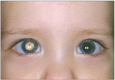
Mechanism
Normal binocular alignment depends on the brain actively using matched, high-quality visual input from both eyes to maintain fusion, the neural process that keeps the two eyes pointed at the same target.
When one eye’s vision is significantly and chronically reduced, whether from a structural, refractive, or pathologic cause, the visual input from that eye becomes less useful for fusion, and the brain has progressively less drive to keep the eye aligned.
Over time, this reduced fusional drive allows the poorly-seeing eye to drift, most commonly into exotropia in an older child or adult, though the specific direction of drift can vary, particularly in younger children where esotropia is also common.
Underlying Causes
- Dense congenital or early childhood cataract, discussed in its own dedicated article on this site, one of the classic pediatric causes of sensory strabismus if not treated promptly
- Corneal opacity or scarring severe enough to significantly degrade the image reaching the retina
- Optic nerve disease, including optic atrophy or a congenital optic nerve anomaly, reducing the quality of visual signal regardless of how clear the eye’s optics are
- Macular or retinal disease, including a retinal scar, significant amblyopia from an untreated refractive error, or a retinal dystrophy affecting central vision
- Any acquired cause of significant monocular vision loss at any age, including trauma, tumor, or vascular events affecting one eye disproportionately
All-fit Slit-Lamp Adapter
Record and share exactly what you see at the slit lamp. One adapter fits any slit lamp or surgical microscope — and any smartphone.
From Choroida — the team behind this siteClinical Presentation
The misalignment itself is usually what brings the patient to medical attention, sometimes noticed by family members well before any underlying visual problem is recognized or reported.
Age at onset of the underlying visual loss significantly affects the pattern: sensory strabismus developing in early childhood, during the critical period of visual development, often shows esotropia, while onset later in childhood or in adulthood more typically produces exotropia.
A careful exam looking specifically for the underlying cause, rather than treating the strabismus as an isolated primary finding, is essential, since the misalignment itself is a downstream consequence rather than the actual disease process.
Evaluation
A complete ocular exam, including assessment of media clarity, a dilated fundus exam, and evaluation of the optic nerve, aims to identify the specific cause of the underlying vision loss driving the strabismus.
Additional testing, including electrophysiology for suspected retinal or optic nerve disease, or neuroimaging when a structural or neurological cause is suspected, may be needed depending on what the initial exam suggests.
In a young child, distinguishing sensory strabismus from a primary strabismus with secondary amblyopia can be genuinely difficult, and both processes may in fact be present and interacting in the same eye.
Imaging of the orbit and brain is warranted when the underlying eye exam does not adequately explain the degree of vision loss, since an intracranial or orbital process affecting the visual pathway can present this way as well.
Management
Treatment is directed first at the underlying cause whenever that cause is treatable, such as cataract extraction or, in a young child, treatment of the underlying refractive or visual problem during the critical period of visual development.
Once the underlying cause has been addressed or found to be untreatable, strabismus surgery can be considered to realign the eyes, primarily for cosmetic purposes in most sensory strabismus cases, since binocular fusion is unlikely to be restored when the underlying vision loss is longstanding or permanent.
Setting expectations accurately with the patient or family before surgery is important, distinguishing the realistic cosmetic goal of straightening the eyes from the generally unrealistic goal of restoring binocular vision in an eye with significant, permanent underlying vision loss.
Recurrent drift after an initially successful alignment surgery is also more common in sensory strabismus than in strabismus with normal or near-normal vision in both eyes, since the same underlying lack of fusional drive that caused the original deviation persists after the surgery corrects the eye position.


Document what you see
Two smartphone imaging tools built for everyday clinic use — one for the slit lamp, one for the fundus.
From Choroida — the team behind this siteReferences
- American Academy of Ophthalmology. Basic and Clinical Science Course, Section 6: Pediatric Ophthalmology and Strabismus.
- von Noorden GK, Campos EC. Binocular Vision and Ocular Motility: Theory and Management of Strabismus.
- Bowling B. Kanski’s Clinical Ophthalmology.
