
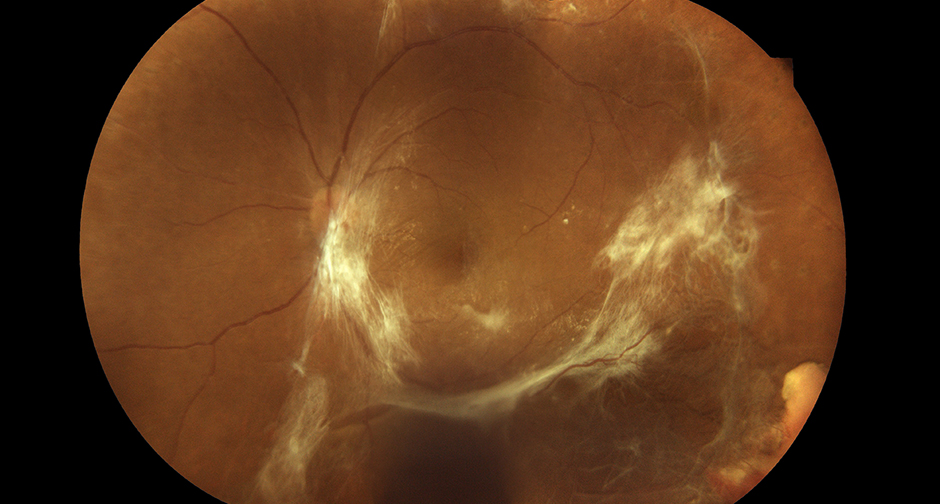
Vitreomacular traction (VMT) occurs when the posterior vitreous cortex remains partially attached to the macula during vitreous separation, pulling on the retinal surface and distorting the foveal contour.
It sits on a spectrum between simple vitreomacular adhesion, which causes no retinal distortion, and full-thickness macular hole, where the retina has torn under sustained traction.
Where a given patient falls on that spectrum, and whether it is actively progressing, is what drives the decision between watching and treating.
Pathophysiology
As the vitreous ages, it liquefies and progressively separates from the retina in a process called posterior vitreous detachment. In most eyes, this separation completes smoothly and the vitreous releases evenly from the retinal surface.
In some eyes, the vitreous cortex remains abnormally adherent over the macula even as it separates elsewhere, and this focal, persistent attachment is what generates traction on the underlying retina.
Sustained traction distorts the foveal contour, can create intraretinal cystic changes, and in more severe cases elevates the fovea away from the retinal pigment epithelium. If traction continues, it can progress to a full-thickness macular hole.
Classification
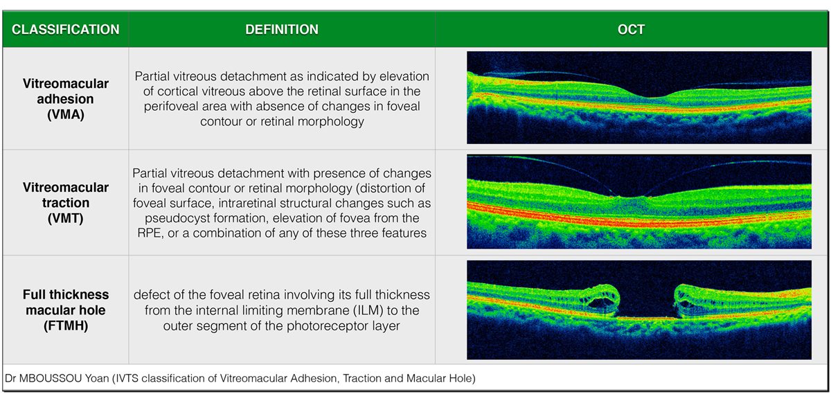
The International Vitreomacular Traction Study Group classification separates this spectrum into three stages based on OCT appearance.
- Vitreomacular adhesion – partial vitreous detachment with persistent perifoveal attachment but no distortion of foveal contour or retinal architecture
- Vitreomacular traction – partial vitreous detachment with clear distortion of foveal contour, intraretinal structural change such as pseudocyst formation, or foveal elevation from the RPE
- Full-thickness macular hole – a defect spanning the complete retinal thickness from the internal limiting membrane to the outer photoreceptor segment
VMT is further described as focal, when the area of persistent adhesion is 1500 microns or less in diameter, or broad, when it exceeds that. This distinction matters because focal adhesion is more likely to release spontaneously than broad adhesion.
Fundus Explorer Pro
Photograph the retinal findings described here with the phone already in your pocket — 22 D optics and built-in illumination in one handheld unit.
From Choroida — the team behind this siteClinical Presentation
Symptoms depend on the degree of traction and whether the fovea itself is distorted. Many cases of mild vitreomacular adhesion are entirely asymptomatic and found incidentally on OCT performed for another reason.
Once traction produces foveal distortion, patients typically report metamorphopsia, blurred or distorted central vision, and sometimes a central or paracentral scotoma. Symptoms can fluctuate as the degree of traction changes over time, occasionally worsening or, less often, improving as the vitreous shifts.
Diagnostic Evaluation
OCT is the essential investigation, both to confirm the diagnosis and to classify it precisely along the adhesion-traction-hole spectrum described above. Spectral-domain OCT clearly demonstrates the vitreous attachment, its width, and any associated retinal structural change.
Amsler grid testing can be used to document and monitor metamorphopsia over time, giving a simple functional correlate to the anatomical OCT findings. Visual acuity should be recorded at baseline and monitored at follow-up to track functional impact alongside the anatomical picture.
Differential Diagnosis
- Epiretinal membrane, which can coexist with VMT but is a distinct fibrocellular proliferation on the retinal surface rather than persistent vitreous attachment
- Lamellar macular hole, a partial-thickness defect with a different OCT signature, often associated with epiretinal membrane rather than vitreous traction
- Cystoid macular oedema from other causes, distinguished by the absence of vitreous attachment and a different underlying mechanism, such as post-cataract surgery inflammation or diabetic retinopathy
- Central serous chorioretinopathy, which can also cause metamorphopsia but shows subretinal fluid rather than traction on OCT
Management
Observation
Asymptomatic vitreomacular adhesion, and mild VMT with good vision and minimal symptoms, are reasonable to observe. Focal adhesion in particular has a meaningful chance of releasing spontaneously over months, resolving the traction without any intervention.
Pharmacologic Vitreolysis
Intravitreal ocriplasmin can enzymatically dissolve the vitreomacular adhesion in selected cases, most effective in eyes with focal adhesion, no epiretinal membrane, and a relatively small macular hole if one is present. Success rates are lower in eyes with broad adhesion or coexisting epiretinal membrane, which limits how broadly this option applies in practice.
Pars Plana Vitrectomy
Surgical removal of the vitreous with membrane peeling is used for symptomatic VMT that fails to release spontaneously or with pharmacologic treatment, and is the standard approach once a full-thickness macular hole has developed. Surgery reliably relieves the mechanical traction, though visual recovery still depends on how long the fovea was distorted before treatment.
Prognosis
Spontaneous release of vitreomacular adhesion, when it occurs, generally leads to full anatomical and visual recovery without further intervention. Focal adhesion is considerably more likely to resolve this way than broad adhesion.
Progression to macular hole occurs in a meaningful minority of untreated VMT cases, particularly those with broad adhesion or coexisting epiretinal membrane, which is why these features are followed more closely rather than simply observed indefinitely.
When treatment, pharmacologic or surgical, is needed, outcomes are generally favourable, with visual improvement in the majority of appropriately selected patients, though the degree of recovery correlates with how long significant foveal distortion was present before the traction was relieved.


Document what you see
Two smartphone imaging tools built for everyday clinic use — one for the slit lamp, one for the fundus.
From Choroida — the team behind this siteReferences
- Duker JS, Kaiser PK, Binder S, et al. The International Vitreomacular Traction Study Group classification of vitreomacular adhesion, traction, and macular hole. Ophthalmology. 2013.
- Stalmans P, Benz MS, Gandorfer A, et al. Enzymatic vitreolysis with ocriplasmin for vitreomacular traction and macular holes. New England Journal of Medicine. 2012.
- Johnson MW. Posterior vitreous detachment: evolution and complications of its early stages. American Journal of Ophthalmology. 2010.
- Vitreomacular Traction. EyeWiki, American Academy of Ophthalmology.
- Vitreomacular Traction Syndrome. StatPearls, NCBI Bookshelf.
Test yourself
A few questions straight from this article.
-
What defines vitreomacular traction?
The posterior vitreous cortex stays partly attached to the macula while separating elsewhere, pulling on the retinal surface. It sits between simple adhesion and full-thickness hole. -
What is the underlying mechanism of vitreomacular traction as the vitreous ages?
In most eyes posterior vitreous detachment completes smoothly. When the cortex stays focally stuck over the macula, that persistent attachment generates the traction. -
On OCT, what separates vitreomacular traction from vitreomacular adhesion?
Adhesion means persistent perifoveal attachment with no distortion at all. Traction adds contour change, pseudocyst formation, or foveal elevation from the RPE. -
Above what adhesion diameter is vitreomacular traction classified as broad?
Adhesion of this width or less is called focal, and anything wider is broad. The distinction matters because focal adhesion is more likely to release on its own. -
What symptom do patients typically report once traction distorts the fovea?
A central or paracentral scotoma may accompany it, and symptoms can fluctuate as traction shifts. Milder adhesion is often found incidentally on OCT done for another reason. -
Which investigation confirms and classifies vitreomacular traction?
Spectral-domain OCT shows the vitreous attachment, its width, and any retinal structural change, which is exactly what the classification depends on. -
How does central serous chorioretinopathy differ from vitreomacular traction on OCT?
Both can present with metamorphopsia, so the OCT appearance is what separates them. The vitreoretinal interface is normal in central serous chorioretinopathy. -
In which eyes is intravitreal ocriplasmin most effective for vitreomacular traction?
Success rates drop markedly with broad adhesion or a coexisting epiretinal membrane, which is what limits how widely this option is useful in practice. -
What is the standard treatment once a full-thickness macular hole has developed?
Surgery reliably relieves the mechanical traction. It is also used for symptomatic traction that fails to release spontaneously or with pharmacologic treatment. -
What most influences visual recovery once vitreomacular traction is relieved?
Outcomes are generally favourable in well-selected patients, but the degree of recovery tracks the duration of significant foveal distortion before traction was released.

Writes and reviews the clinical articles on Educate. Every post is checked against peer-reviewed sources before it goes live.


