
CASE REPORT
A 65-year-old woman came to a medical Centre in March 2013 with symptoms of blurred vision in her left eye. She had a history of branch retinal vein occlusion in the left eye in January 2012 and 2 sessions of retinal laser photocoagulation in the upper quadrants.

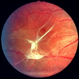
On examination, the best corrected visual acuity (BCVA) was 0.7 in the left eye. The lens was clear and the fundus examination showed pigment atrophy and retinal wrinkling in the macular area, occlusion of the upper-temporal branch of the central retinal vein, traces of laser photocoagulation in the upper quadrants, and peripheral retinal neovascularization with focal vitreous hemorrhages.
Optical coherence tomography (OCT) revealed incomplete posterior vitreous detachment, ERM on the macular surface, and slight diffuse retinal edema. The diagnosis was Epiretinal Membrane (ERM).
Epiretinal Membrane DISEASE entity
Epiretinal Membrane (ERM) is a fibrocellular tissue found on the inner surface of the retina. It is semi-translucent and proliferates on the surface of the internal limiting membrane.

Idiopathic ERMs are the most common presentation. Secondary ERMs occur in association with retinal vascular diseases including diabetic retinopathy, retinal vein occlusion, ocular inflammatory disease, trauma, intraocular surgery, intraocular tumors, and retinal tear or detachment.
Other risk factors include age, posterior vitreous detachment, and history of Epiretinal Membrane (ERM) in the fellow eye.
The mean age of ERM diagnosis is 65 years old. The incidence of developing an ERM in the primary eye is 1.1% per year. The incidence of developing an ERM in the fellow eye is 2.7% per year.
Retinal glial and retinal pigment epithelial cells are the major components. Fibrous astrocytes, fibrocytes, myofibrocytes, and macrophages can also be identified in the pathological analysis.
Fundus Explorer Pro
Photograph the retinal findings described here with the phone already in your pocket — 22 D optics and built-in illumination in one handheld unit.
From Choroida — the team behind this siteEpiretinal Membrane Diagnosis
A clinical diagnosis is based on history and clinical exam, including slit lamp and dilated fundus examination. In some cases, Optical Coherence Tomography (OCT) is useful in the diagnosis, quantification of retinal thickness, and management of this condition.
MANAGEMENT of Epiretinal Membrane
General treatment
The most important issue in the management of idiopathic ERMs is the presence of visual complaints. Visual symptoms can be variable and sometimes independent of clinical severity.
Surgery
Epiretinal Membrane (ERM) surgery is the most common vitreoretinal surgery performed as reported by the Centers for Medicare and Medicaid Services.
Surgery involves a pars plana vitrectomy procedure with membrane peel. A number of different instruments can be used to facilitate removal including intraocular forceps, pick, diamond dusted instruments, as well as other instruments.
In many cases, internal limiting membrane peel is also performed concurrently with Epiretinal Membrane (ERM) removal.

Surgical follow-up
Surgery is indicated if the patient has significant visual complaints of visual decline and /or metamorphopsia.
The follow-up is similar for most eyes following pars plana vitrectomy surgery. Visual acuity improvement does not occur immediately in some patients.
This is highly dependent on preoperative characteristics, duration of the Epiretinal Membrane, as well as other factors. Most patients improve by 3-6 months postoperatively. However, some may experience improvement 1-2 years postoperatively.


Document what you see
Two smartphone imaging tools built for everyday clinic use — one for the slit lamp, one for the fundus.
From Choroida — the team behind this siteREFERENCES
- Fraser-Bell S, Guzowski M, Rochtchina E, et al. Five-year cumulative incidence and progression of epiretinal membranes: the Blue Mountains Eye Study. Ophthalmology 2003;110(1):34-40.
- Gillis K. Medicare Physician Payment Schedule Services for 2001 – A Summary of Claims Data. In: Physician Marketplace Report. Chicago: American Medical Association, 2003.
- Gupta OP, Brown GC, Brown MM. A value-based medicine cost-utility analysis of idiopathic epiretinal membrane surgery. Am J Ophthalmol 2008;145(5):923-8.
Test yourself
A few questions straight from this article.
-
Where does an epiretinal membrane proliferate?
An epiretinal membrane is semi-translucent fibrocellular tissue that proliferates on the inner retinal surface, over the internal limiting membrane. -
Which form of epiretinal membrane is the most common presentation?
Idiopathic epiretinal membranes are the most common presentation; secondary membranes follow retinal vascular disease, inflammation, trauma, surgery, tumours or retinal breaks. -
Which cells are the major components of an epiretinal membrane?
Retinal glial and RPE cells predominate, with fibrous astrocytes, fibrocytes, myofibrocytes and macrophages also identified on pathological analysis. -
What is the mean age at which epiretinal membrane is diagnosed?
The mean age of epiretinal membrane diagnosis is 65 years, and increasing age is itself a risk factor. -
What is the annual incidence of developing an epiretinal membrane in the fellow eye?
The fellow eye develops an epiretinal membrane at 2.7% per year, compared with 1.1% per year in the primary eye. -
Which retinal vascular disorder is associated with secondary epiretinal membrane formation?
Secondary epiretinal membranes arise with retinal vascular diseases such as diabetic retinopathy and retinal vein occlusion, as well as inflammation, trauma and surgery. -
Beyond confirming the diagnosis, what does OCT add in epiretinal membrane assessment?
Diagnosis rests on history plus slit lamp and dilated fundus examination, with OCT useful for confirming the membrane, quantifying retinal thickness and guiding management. -
Which finding indicates surgery for a patient with epiretinal membrane?
The decisive factor is symptoms: surgery is indicated when the patient reports significant visual decline or metamorphopsia, since symptoms can be independent of clinical severity. -
What surgical procedure is performed for a symptomatic epiretinal membrane?
Surgery is pars plana vitrectomy with membrane peel using forceps, picks or diamond-dusted instruments, and in many cases an internal limiting membrane peel is done concurrently. -
When do most patients show visual improvement after epiretinal membrane surgery?
Visual recovery is not immediate. Most patients improve by 3 to 6 months, though some continue to gain vision 1 to 2 years after surgery.
