
Gelatinous drop-like corneal dystrophy produces a dramatic, elevated, mulberry-shaped subepithelial mass, and its rarity combined with a presentation that can initially resemble chronic allergic or inflammatory keratoconjunctivitis means this diagnosis is genuinely easy to delay, sometimes for years, unless a clinician specifically considers a primary amyloid-depositing corneal dystrophy in a patient with a stubbornly recurrent, gelatinous-appearing corneal mass.
What’s Actually Accumulating
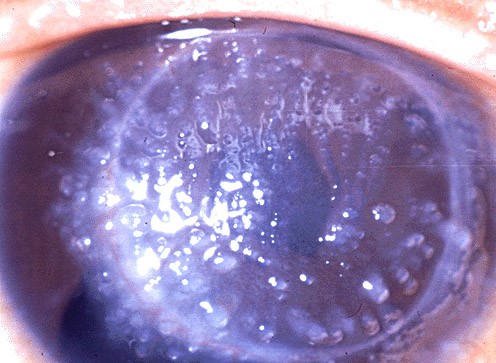
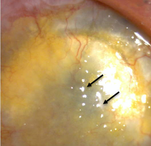
This dystrophy results from progressive, severe subepithelial deposition of amyloid, producing masses that protrude prominently from the corneal surface, distinguishing it in both severity and appearance from the more localized, generally flatter amyloid pattern of lattice corneal dystrophy.
Genetics
Gelatinous drop-like corneal dystrophy is inherited in an autosomal recessive pattern and results from mutations in the TACSTD2 gene (also known as M1S1), which encodes a protein important for normal corneal epithelial barrier function; loss of this barrier function is thought to allow abnormal amyloid deposition to accumulate progressively beneath the epithelium.
All-fit Slit-Lamp Adapter
Record and share exactly what you see at the slit lamp. One adapter fits any slit lamp or surgical microscope — and any smartphone.
From Choroida — the team behind this siteClinical Presentation
- Onset is typically in the first or second decade of life, presenting initially with milder symptoms, including photophobia, tearing, and foreign body sensation, that can be mistaken for a chronic, nonspecific keratoconjunctivitis
- Progressive development of elevated, gelatinous, mulberry-shaped or nodular subepithelial masses, which can become quite prominent and are the hallmark, ultimately unmistakable finding once sufficiently developed
- Significant visual impairment develops as the masses enlarge and multiply, obstructing the visual axis
- Bilateral, and typically progressive over years if untreated
- A notably high rate of recurrence after surgical removal is a defining, frustrating feature of this condition, related to the ongoing underlying epithelial barrier dysfunction that continues to drive amyloid deposition even after visible masses are excised
Why Early Recognition Matters
Because early disease can resemble a chronic, nonspecific inflammatory or allergic conjunctivitis-keratitis picture, and because this is a rare condition many clinicians will encounter only once or never in a career, delayed diagnosis is common, and patients may be treated for presumed allergic or inflammatory disease for a prolonged period before the characteristic elevated, gelatinous masses become unmistakable.
A geographic clustering has been described, with higher prevalence reported in certain populations, including parts of Japan, which can inform the index of suspicion depending on patient background, though the condition occurs worldwide.
Diagnostic Evaluation
- Slit-lamp examination identifying the characteristic elevated, gelatinous, mulberry-shaped subepithelial mass, which becomes increasingly distinctive as the disease progresses
- A family history consistent with autosomal recessive inheritance, and parental consanguinity in some populations, given the recessive pattern
- Biopsy or examination of surgically excised tissue, showing amyloid deposition on histopathology with appropriate staining, confirms the diagnosis definitively
- Genetic testing for TACSTD2 mutations supports diagnosis and family counseling
Management
Surgical Excision
Surgical removal of the amyloid masses, whether by superficial keratectomy, lamellar keratoplasty, or, for more extensive or recurrent disease, penetrating keratoplasty, is the primary treatment for visually significant disease.
The Recurrence Challenge
This condition is notorious for a high rate of recurrence after surgical treatment, sometimes within just a few years, reflecting the persistent underlying epithelial barrier defect that continues to drive amyloid formation in the patient’s own corneal tissue regardless of the surgical approach used, which is an essential point for realistic patient counseling before any surgical intervention.
- Some reports suggest that more extensive excision, including lamellar or penetrating keratoplasty rather than superficial keratectomy alone, may offer somewhat longer recurrence-free intervals, though recurrence remains a significant long-term concern with any surgical approach
Adjunctive Measures
Soft contact lens wear has been explored as a way to mechanically protect the ocular surface and potentially slow recurrence in some reported cases, though this remains a secondary, adjunctive strategy rather than a primary treatment.
Recurrence after surgery
A hallmark of this dystrophy is the return of amyloid deposits in the graft, sometimes within a few years of keratoplasty. Superficial keratectomy and lamellar keratoplasty are often repeated, and some patients are managed with a contact lens or a keratoprosthesis in advanced cases. Because the disease affects the surface, procedures that remove only the diseased layers may be preferable to full-thickness grafts.
Counselling the patient
Tell patients that surgery improves vision for a period but is not a cure. Sun protection, lubrication, and regular follow-up delay problems, and early treatment of erosions and new vessels helps. Families should be advised about the recessive pattern of inheritance, and genetic counselling should be offered when relatives are at risk.
Prognosis
Gelatinous drop-like corneal dystrophy is one of the more challenging corneal dystrophies to manage definitively, given its high recurrence rate after surgery and its progressive, bilateral nature.
Patients require realistic counseling about the likelihood of needing repeated surgical intervention over their lifetime, and sustained, long-term ophthalmic follow-up is essential given both the recurrence risk and the potential for progressive visual impairment if the condition is not actively monitored and managed.


Document what you see
Two smartphone imaging tools built for everyday clinic use — one for the slit lamp, one for the fundus.
From Choroida — the team behind this siteReferences
- Tsujikawa M, Kurahashi H, Tanaka T, et al. Identification of the gene responsible for gelatinous drop-like corneal dystrophy. Nat Genet. 1999;21:420-423.
- Weiss JS, Moller HU, Aldave AJ, et al. IC3D classification of corneal dystrophies–edition 2. Cornea. 2015;34:117-159.
- Ide T, Nishida K, Maeda N, et al. A spectrum of clinical manifestations of gelatinous drop-like corneal dystrophy in Japan. Am J Ophthalmol. 2004;137:1081-1084.
- Kinoshita S, Nishida K, Dota A, Saito I, Yokoi N. Epithelial barrier function and ultrastructure of gelatinous drop-like corneal dystrophy. Cornea. 2000;19:551-555.
