
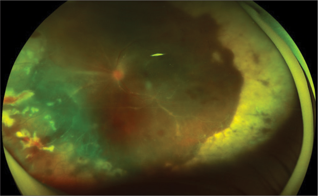
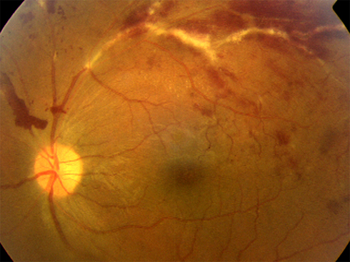
Retinal vasculitis is inflammation of the retinal vessels, recognized by perivascular sheathing, vascular leakage on angiography, and often vascular occlusion.
It is a clinical sign and not a diagnosis, and the underlying cause may be infectious, systemic inflammatory, or limited to the eye.
Treating an unrecognized infection with corticosteroids can worsen the disease, so the workup comes before immunosuppression.
A pattern-based approach shortens the list of possible causes and guides the tests that need to be ordered.
What Is Retinal Vasculitis?
Retinal vasculitis describes inflammation involving retinal arterioles, venules, or both.
It may occur as part of uveitis or as an isolated finding.
Clinical signs include:
- Perivascular sheathing or cuffing
- Vascular leakage and staining on fluorescein angiography
- Retinal hemorrhages and cotton wool spots
- Vascular occlusion with capillary nonperfusion
- Retinal or optic disc neovascularization
- Vitritis and macular edema
Fluorescein angiography, preferably widefield, is the most informative test, because it shows leakage and nonperfusion that are not visible on ophthalmoscopy.
Classification by Vessel Involved
Periphlebitis
Venous involvement is the most common pattern.
It is seen in sarcoidosis, tuberculosis, multiple sclerosis, Behçet disease, Eales disease, and pars planitis.
Arteritis
Arterial involvement suggests systemic vasculitis, lupus, toxoplasmosis, acute retinal necrosis, syphilis, cat scratch disease, and Susac syndrome (see Susac syndrome).
Severe arteritis may lead to branch or central retinal artery occlusion.
Occlusive Versus Nonocclusive
Occlusive disease carries a higher risk of ischemia and neovascularization, and it needs closer attention.
Fundus Explorer Pro
Photograph the retinal findings described here with the phone already in your pocket — 22 D optics and built-in illumination in one handheld unit.
From Choroida — the team behind this siteCauses
Infectious
- Tuberculosis (see ocular tuberculosis)
- Syphilis (see ocular syphilis)
- Toxoplasmosis
- Viral retinitis, including herpes viruses and cytomegalovirus (see acute retinal necrosis)
- Bartonella, Lyme disease, brucellosis, and other bacterial infections
Noninfectious Systemic
- Behçet disease
- Sarcoidosis (see ocular sarcoidosis)
- Systemic lupus erythematosus and antiphospholipid syndrome
- Granulomatosis with polyangiitis and polyarteritis nodosa
- Multiple sclerosis
- Inflammatory bowel disease
Ocular-Limited
- Idiopathic retinal vasculitis, often with aneurysms and neuroretinitis (see IRVAN)
- Frosted branch angiitis (see frosted branch angiitis)
- Hemorrhagic occlusive retinal vasculitis after intracameral or intravitreal drugs (see HORV)
- Birdshot chorioretinopathy (see birdshot chorioretinopathy)
Some patients remain undiagnosed after a full workup, and idiopathic retinal vasculitis is a diagnosis of exclusion.
Diagnostic Workup
The initial workup should be guided by the pattern, the patient’s geography, and the systemic review.
A common screening panel includes:
- Complete blood count, ESR, CRP
- Treponemal and nontreponemal syphilis serology
- Tuberculosis testing (interferon-gamma release assay or tuberculin skin test) and chest imaging
- Serum ACE, lysozyme, and chest CT if sarcoidosis is suspected
- ANA, ANCA, antiphospholipid antibodies, and complement when lupus or systemic vasculitis is possible
- HIV testing
- Toxoplasma serology when the pattern fits
- HLA-B51 and clinical criteria in suspected Behçet disease
- MRI of the brain and orbits when demyelination or Susac syndrome is a concern
Vitreous or aqueous sampling with PCR for herpes viruses and Toxoplasma is helpful when the diagnosis is unclear or the disease is severe.
Widefield fluorescein angiography defines the extent of leakage and nonperfusion and guides laser therapy.
OCT identifies macular edema, and OCT angiography shows capillary dropout.
Angiographic Patterns That Help
Certain angiographic patterns narrow the differential before the serology returns.
- Diffuse capillary leakage with a fern-like pattern and disc leakage suggests Behçet disease
- Peripheral venous leakage with nonperfusion and neovascularization in a young patient suggests Eales disease or tuberculosis
- Arteriolar wall staining and segmental occlusion suggests systemic vasculitis, syphilis, or acute retinal necrosis
- Multiple aneurysmal dilations of the arterioles at bifurcations with disc leakage suggests IRVAN
- A confluent, frosted appearance with diffuse sheathing suggests frosted branch angiitis, often viral or lymphomatous
- Predominantly peripheral leakage with snowbanking suggests intermediate uveitis, sarcoidosis, or multiple sclerosis
Patterns are guides and not proof, and overlap is frequent.
Common Pitfalls
A few errors recur in practice.
- Starting corticosteroids before excluding tuberculosis, syphilis, and viral retinitis
- Assuming the vasculitis is idiopathic when a systemic workup has not been completed
- Missing peripheral ischemia because only central fields were imaged
- Treating macular edema and neovascularization but not the underlying inflammation
- Overlooking lymphoma in an older patient with vitritis and a frosted vascular appearance
Repeated review is needed because the diagnosis may become apparent only as the disease evolves.
Management
Treat the Cause
Infectious causes need specific antimicrobial therapy.
Corticosteroids should be started only after the appropriate antimicrobial cover is in place, and after infection has been reasonably excluded.
Corticosteroids
Oral prednisone, pulse intravenous methylprednisolone, and periocular or intravitreal injections control acute inflammation.
They are not suitable for long-term use because of systemic and ocular side effects.
Steroid-Sparing Agents
Chronic noninfectious disease usually needs immunomodulatory therapy.
Options include azathioprine, mycophenolate, methotrexate, cyclosporine, interferon alpha, and biologics.
In noninfectious intermediate, posterior, and panuveitis, adalimumab reduced the risk of treatment failure in the VISUAL I and VISUAL II trials, and infliximab is used in refractory Behçet-related disease.
Ischemia and Neovascularization
Panretinal photocoagulation to nonperfused retina reduces the drive for neovascular growth.
Anti-VEGF injections help neovascularization and macular edema, although they do not treat the inflammation.
Vitrectomy is used for nonclearing vitreous hemorrhage and tractional detachment.
Prognosis
Outcome depends on the cause, on how early it is treated, and on whether ischemia develops.
Recurrences are common in chronic disease, and follow-up with angiography helps detect silent activity.
Some patients relapse when treatment is tapered, which is why regular review continues for years.


Document what you see
Two smartphone imaging tools built for everyday clinic use — one for the slit lamp, one for the fundus.
From Choroida — the team behind this siteReferences
- Rosenbaum JT, Sibley CH, Lin P. Retinal vasculitis. Curr Opin Rheumatol. 2016;28:228-235.
- Graham EM, Stanford MR, Sanders MD, Kasp E, Dumonde DC. A point prevalence study of 150 patients with idiopathic retinal vasculitis: 1. Diagnostic value of ophthalmological features. Br J Ophthalmol. 1989;73:714-721.
- Jaffe GJ, Dick AD, Brezin AP, et al. Adalimumab in patients with active noninfectious uveitis. N Engl J Med. 2016;375:932-943.
- Nguyen QD, Merrill PT, Jaffe GJ, et al. Adalimumab for prevention of uveitic flare in patients with inactive non-infectious uveitis controlled by corticosteroids (VISUAL II): a multicentre, double-masked, randomised, placebo-controlled phase 3 trial. Lancet. 2016;388:1183-1192.
- Ali A, Ku JH, Suhler EB, Choi D, Rosenbaum JT. The course of retinal vasculitis. Br J Ophthalmol. 2014;98:785-789.
