
CASE REPORT
A 64-year-old man experienced a worsening vision for 2 years. His best-corrected visual acuity was worse than 20/30 OU since he was 20 years old, and he has not been permitted to have a driver’s license.

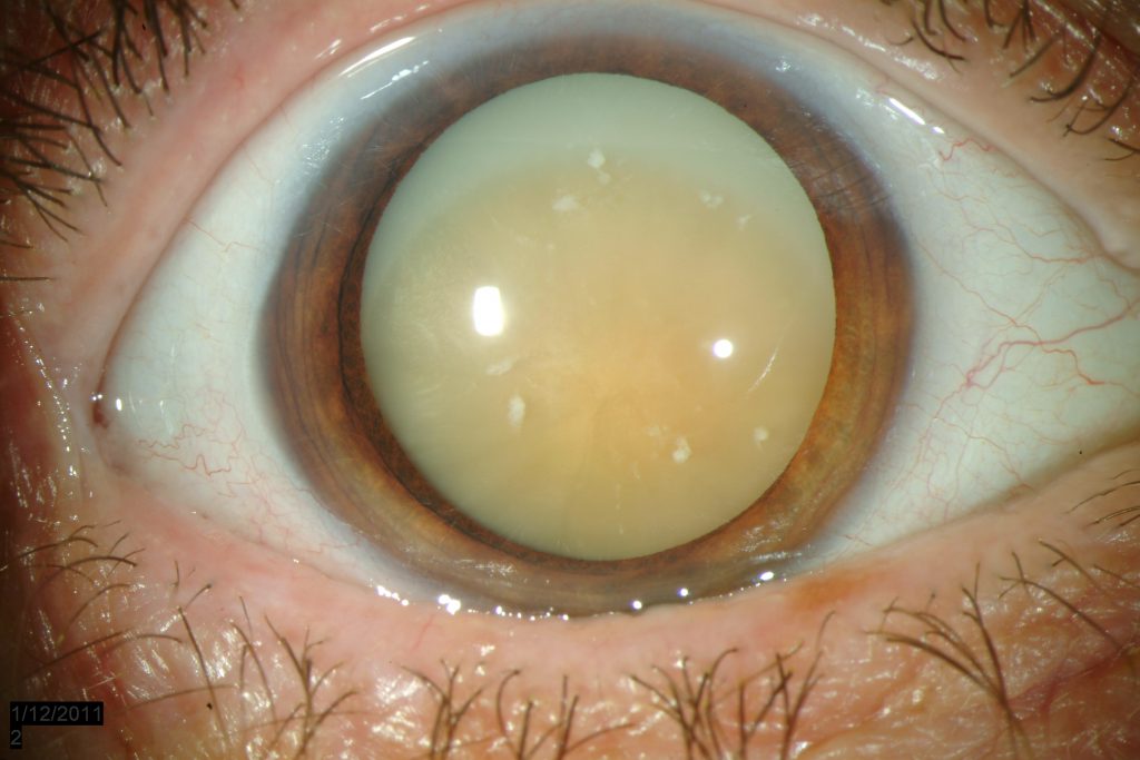
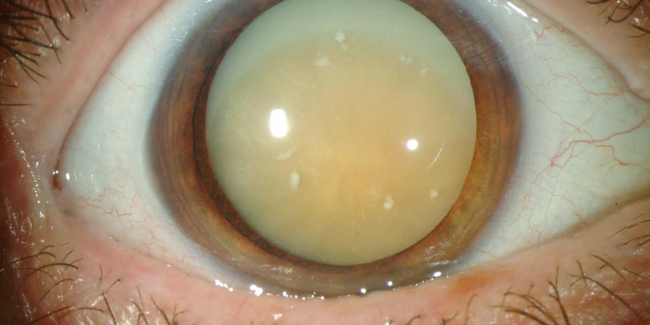
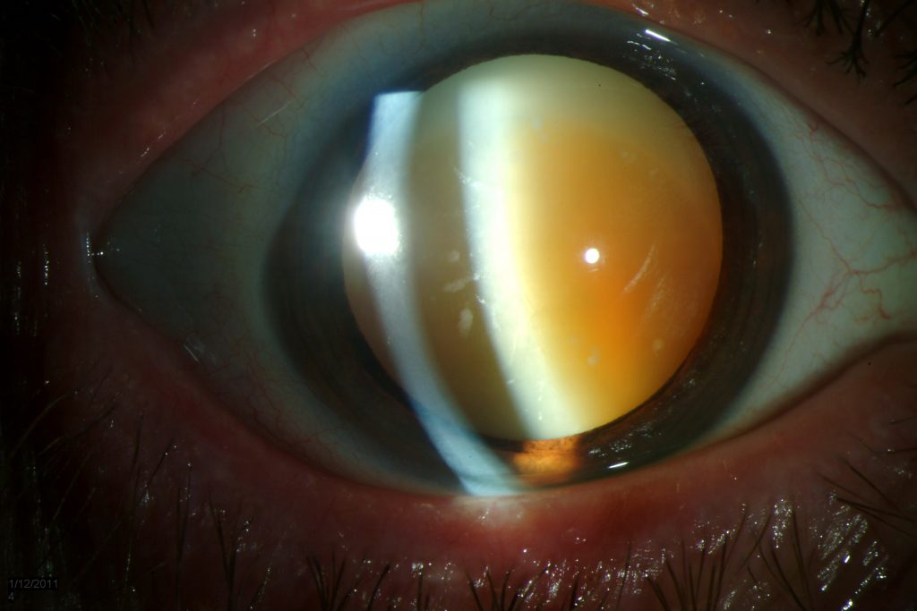
On the initial examination, his best-corrected visual acuity was 20/300 OD and 20/60 OS. Slitlamp biomicroscopy revealed a solid nucleus that descended to the lower equatorial region and a round, isolated posterior capsular opening that was seen through the hydrated cortex in his right eye.
There was no iridodonesis, and the intraocular pressure was normal. His left eye had a cataractous lens with an opacity of the posterior pole.
The slitlamp appearance of the right eye shows a cataract with an isolated posterior capsular opening (arrow). Slitlamp appearance of the left eye shows posterior polar cataract.
Fundus photograph of the right eye showing crystal-like foamy substances on the macular region. The slitlamp appearance of the right eye 1 week after cataract surgery. The intraocular lens was well centered.
The patient was diagnosed as having morgagnian cataract
DISEASE
Morgagnian cataract is a form of hypermature cataract formed by liquefaction of the cortex and sinking of the dense nucleus to the bottom of the capsular bag.
The name “Morgagnian” derives from Giovanni Battista Morgagni, the 18th-century anatomical pathologist.
MANAGEMENT
Surgical removal of the cataractous lens followed by intraocular lens implantation is the treatment modality of choice.
Surgery
- Extracapsular Cataract Extraction (ECCE), Small incision cataract surgery (SICS), or phacoemulsification can be performed for the cataract extraction. This is followed by intraocular lens implantation (posterior chamber intraocular lens (PCIOL )/ anterior chamber intraocular lens (ACIOL).
- Intraocular lens scaffold technique has been described to prevent posterior capsule rupture for phacoemulsification of a Morgagnian cataract. This technique uses the IOL as a scaffold to prevent the vulnerable posterior capsule from rupturing during nuclear emulsification in Morgagnian cataract.
The technique prevents rupture of the floppy posterior capsule by providing constant support to it. The scaffold provides stable inflation of the capsular bag and prevents inadvertent emulsification.
Concurrently, it prevents the dehiscence of weak zonular fibers by minimizing the stress on the zonular apparatus.
HOW TO TAKE SLIT-LAMP EXAM IMAGES BY A SMARTPHONE?
Smartphone slit-lamp photography is the new advancement in the field of science and technology in which the photographs of the desired slit-lamp finding can be taken with smartphones by using the slit-lamp adaptors.

REFERENCES
- Basic Clinical Science Course (BCSC) of the American Academy of Ophthalmology. Section 11. 2014 – 2015.
- Rozenbaum I. The Faces Behind the Eponyms. Arch Ophthalmol. 2008;126(6):846. doi:10.1001/archophthalmol.2007.63
- Bron AJ, Habgood JO. Morgagnian cataract. Trans Ophthalmol Soc U K. 1976 Jul;96(2):265-77.
- Roger F. Steinert. Cataract surgery 3rd edition. Chapter 1, The pathology of cataracts. CA, USA: Elsevier Inc.; 2010
- Deshmukh S, Bhattacharjee H, Gupta K. “Triangle sign”in Morgagnian cataract. Indian J Ophthalmol 2019;67:137.
- KAUFMAN SI. Morgagnian cataracts and their complications: with report of a case of spontaneous rupture of the lens capsule causing secondary glaucoma. Archives of Ophthalmology. 1933 Jan 1;9(1):56-63.
Read more: Rosette-Shaped Cataract, Christmas tree cataract, Choroidal Folds,Cerulean Cataract, Best 5 ways for retinal photography.
