
DISEASE
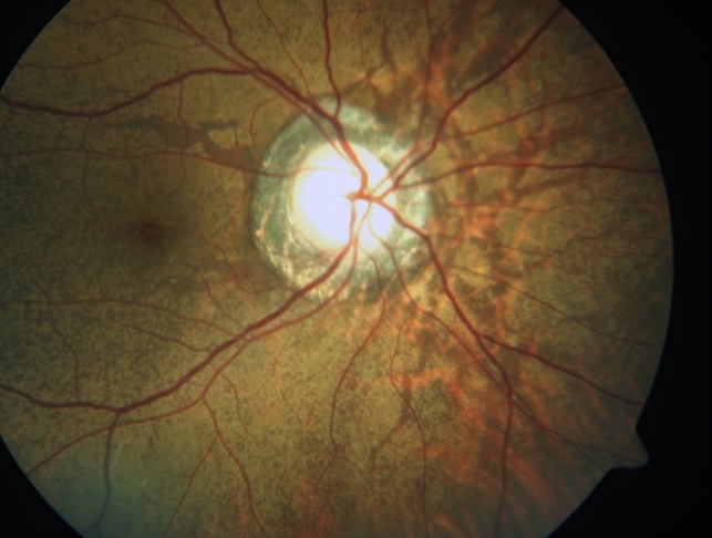
Angioid streaks are bilateral, narrow, irregular lines deep to the retina configured in a radiating fashion emanating from the optic disc, which result from breaks in a weakened Bruch’s membrane.

Ophthalmologists should be aware that there are numerous systemic diseases associated with angioid streaks, the most common being pseudoxanthoma elasticum. The diagnosis of angioid streaks is one that can usually be made clinically.
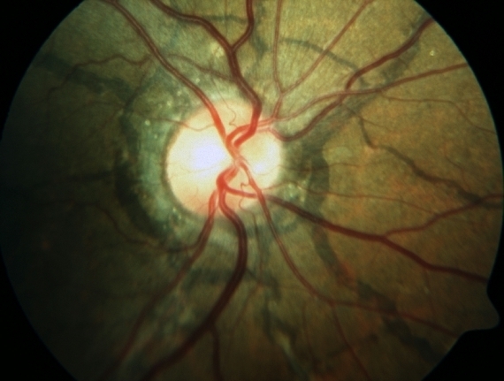
Most patients are asymptomatic but secondary complications can result in choroidal neovascularization, lead to vision loss, and warrant treatment.
Secondary Complications
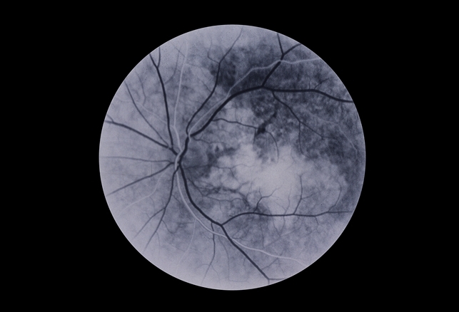
Angioid streaks can cause subretinal hemorrhages from choroidal fibrovascular ingrowth. These hemorrhages can resolve spontaneously or can lead to choroidal neovascularization, which can be recurrent and lead to progressive visual loss.
In fact, recent studies have revealed that in patients with pseudoxanthoma elasticum, the longer the angioid streak, the higher the risk of choroidal neovascularization and macular atrophy.
MANAGEMENT
A general management strategy includes determining if any underlying systemic association is present and if any secondary ocular complication exists that warrants treatment.
Medical Workup
A patient may be sent to the ophthalmologist with a known systemic disease association but at other times may be newly diagnosed with angioid streaks and have no known underlying systemic cause.
The medical workup should be tailored to the individual after a thorough review of the systems is undertaken.
The three most common systemic associations are pseudoxanthoma elasticum, Paget’s disease of bone, and sickle cell hemoglobinopathies, and therefore any screening workup should focus on these entities.
Patients should receive a complete physical examination by an internist, a skin biopsy, and blood testing to include serum alkaline phosphatase, serum calcium and phosphate, and hemoglobin electrophoresis.
Treatment
Angioid streaks are usually asymptomatic requiring observation only. When existent treatment methods can simply restrict the pathology;
however, it cannot be perpetually eliminated. When associated choroidal neovascularization is present, laser photocoagulation and photodynamic therapy have been used in the past with relatively poor results and frequent recurrences.
Intravitreal anti-VEGF treatment is the mainstay of treatment with good results. A combination of photodynamic therapy and bevacizumab has also been shown to regress choroidal neovascularization.
Furthermore, early intervention has been shown to be associated with significantly better outcomes.
Would you have interest in taking retina images by smartphone?
Fundus photography is superior to fundus analysis as it enables intraocular pathologies to be photo captured and encrypted information to be shared with colleagues and patients.
Recent technologies allow smartphone-based attachments and integrated lens adaptors to transform the smartphone into a portable fundus camera and Retinal imaging by smartphone.
RETINAL IMAGING BY YOUR SMARTPHONE
REFERENCES
- Chatziralli I; Saitakis G; Dimitriou E; Chatzirallis A; Stoungioti S; Theodossiadis G; Theodossiadis P; Retina (Philadelphia, Pa.), U.S. National Library of Medicine, Jan. 2019,
- Clarkson JG, Altman RD. Angoid Streaks. Surv Ophthalmol. 1982;26(5)235-46.
- Mahroo OA, Hykin PG Syndrome. “Confirmation That Angioid Streaks Are Not Common in Ehlers-Danlos Syndrome.” JAMA Ophthalmology. 2019 Dec 1;137(12):1463. doi: 10.1001/jamaophthalmol.2019.2549.PMID: 31556922
- Regillo C, et al. Retina and Vitreous (Basic and Clinical Science Course). San Francisco, CA: American Academy of Ophthalmology; 2008:93-4.
- Risseeuw, Sara. “The Extent of Angioid Streaks Correlates With Macular Degeneration in Pseudoxanthoma Elasticum.” American Journal of Ophthalmology, 20 July 2020, www.ajo.com/article/S0002-9394(20)30379-2/fulltext#relatedArticles.
- Singman EL, Doyle JJ. JAMA Ophthalmology. 2019 Mar 1;137(3):239. doi: 10.1001/jamaophthalmol.2018.5995.PMID: 30543351
- Yanoff M, Duker JS. Ophthalmology. 2nd edition. St. Louis, MO: Mosby; 2004:969-72.
- Yanuzzi, LA, The Retinal Atlas. Elsevier; 2010:516-8.
