
Optic nerve sheath fenestration is a surgical procedure that creates small windows in the dural sheath surrounding the optic nerve, relieving elevated cerebrospinal fluid pressure specifically around the nerve and protecting it from the progressive damage that untreated papilledema, discussed in its own dedicated article on this site, can cause.
It addresses the optic nerve locally rather than treating the underlying elevated intracranial pressure systemically, which is the key distinction from cerebrospinal fluid diversion procedures such as a shunt, and it is this local, vision-protective focus that defines the procedure’s specific role in managing idiopathic intracranial hypertension, discussed in its own dedicated article on this site.
Understanding when a local, optic-nerve-focused procedure is preferred over a systemic pressure-diversion approach is central to understanding how these two very different treatment strategies fit together in managing chronically elevated intracranial pressure.
The procedure is performed relatively infrequently overall, reserved specifically for the subset of patients whose papilledema has become or is at serious risk of becoming vision-threatening despite appropriate medical management.
Rationale
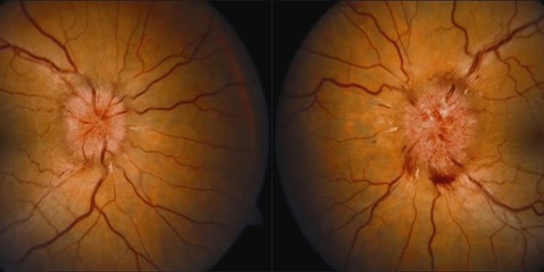
The optic nerve sheath is contiguous with the subarachnoid space surrounding the brain, meaning cerebrospinal fluid pressure elevation is directly transmitted along the nerve sheath, contributing to the axoplasmic flow stasis and disc swelling that defines papilledema.
Creating small windows, or fenestrations, in the sheath allows some of this pressure to be released locally around the nerve, without requiring or achieving any meaningful reduction in intracranial pressure elsewhere in the cranial vault.
This localized mechanism is precisely why the procedure is specifically indicated for protecting vision from papilledema-related optic nerve damage, rather than as a general treatment for the headache and other symptoms caused by elevated intracranial pressure throughout the rest of the cranial cavity.
Indications
- Progressive visual field loss or declining visual acuity from papilledema despite maximal medical management of the underlying elevated intracranial pressure
- Fulminant idiopathic intracranial hypertension with rapid, severe visual decline, where vision protection needs to happen urgently rather than waiting for medical therapy or weight loss measures to take full effect
- Patients who cannot tolerate or have failed medical therapy for idiopathic intracranial hypertension, and for whom the primary concern is specifically protecting vision rather than addressing headache or other systemic symptoms
Fundus Explorer Pro
Photograph the retinal findings described here with the phone already in your pocket — 22 D optics and built-in illumination in one handheld unit.
From Choroida — the team behind this siteOptic Nerve Sheath Fenestration Versus CSF Shunting
Cerebrospinal fluid shunting procedures, including ventriculoperitoneal or lumboperitoneal shunts, reduce intracranial pressure systemically, addressing both the papilledema and other symptoms of elevated pressure, including the often debilitating headache that frequently accompanies idiopathic intracranial hypertension.
Optic nerve sheath fenestration addresses the optic nerve specifically and reliably, but has a less consistent and less direct effect on headache and other systemic symptoms of elevated intracranial pressure, since it does not reduce pressure throughout the broader cranial vault.
The choice between these two approaches, or a combination of both, depends on which symptom, vision loss specifically or the fuller symptom complex of elevated intracranial pressure, is the dominant clinical concern driving the need for surgical intervention in a given patient.
Close collaboration between ophthalmology and neurosurgery is standard when deciding between these options, since neither specialty alone typically holds the full picture needed to weigh vision protection against the broader symptom burden a patient is experiencing.
Technique
The optic nerve is approached surgically, most commonly through a medial orbital approach, to expose the nerve sheath posterior to the globe without disturbing the nerve fibers themselves.
Small linear or window-shaped incisions are made in the dural sheath, allowing cerebrospinal fluid to escape locally and reducing the pressure transmitted to the optic nerve itself.
The procedure can be performed unilaterally, with a documented, though inconsistent, protective effect sometimes observed in the untreated fellow eye as well, or bilaterally when both eyes show significant threat from papilledema.
Outcomes and Complications
Vision stabilization or improvement is achieved in a meaningful proportion of appropriately selected patients, particularly when the procedure is performed before severe, established optic atrophy has already occurred.
Complications include transient or, less commonly, permanent diplopia from manipulation near the extraocular muscles during the surgical approach, along with the general surgical risks inherent to any orbital procedure performed in close proximity to the optic nerve.
The effect of fenestration can wane over time in some patients, as the created windows partially close or scar, occasionally necessitating repeat intervention if papilledema and visual threat recur.
Ongoing monitoring of the optic nerve and visual function after surgery remains important regardless of the initial result, since the underlying driver of elevated intracranial pressure typically persists and continues to require its own separate, ongoing management.


Document what you see
Two smartphone imaging tools built for everyday clinic use — one for the slit lamp, one for the fundus.
From Choroida — the team behind this siteReferences
- American Academy of Ophthalmology. Basic and Clinical Science Course, Section 5: Neuro-Ophthalmology.
- Friedman DI, Liu GT, Digre KB. Revised diagnostic criteria for the pseudotumor cerebri syndrome in adults and children. Neurology.
- Spoor TC, McHenry JG. Long-term effectiveness of optic nerve sheath decompression for pseudotumor cerebri. Archives of Ophthalmology.
