
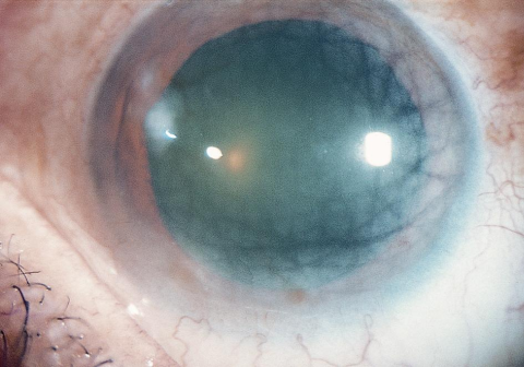
Crocodile shagreen is one of those slit-lamp findings that looks alarming but almost never threatens vision.
It appears as a mosaic of greyish-white polygonal opacities in the corneal stroma, separated by clearer spaces, closely resembling the tanned hide of a crocodile.
The finding is usually bilateral, symmetric, and central, and it is most often discovered incidentally during a routine examination.
For clinicians, the main value of recognizing crocodile shagreen is knowing what it is not. It is easily confused with visually significant corneal dystrophies, yet it rarely requires any intervention.
Understanding its two forms, its associations, and its close mimics helps avoid unnecessary investigation and reassures the patient with confidence.
What Is Crocodile Shagreen?
Crocodile shagreen is a bilateral corneal degeneration characterized by polygonal grey-white opacities with indistinct margins, separated by relatively clear intervening zones.
The pattern gives the cornea a cracked, mosaic appearance likened to crocodile skin.
Key features include:
- Bilateral and broadly symmetric distribution
- Central or paracentral location
- Polygonal opacities with faint, indistinct borders
- Clear spaces between the opacities
- A non-inflammatory, degenerative nature
It is a descriptive clinical entity rather than a single disease, and the same pattern can arise at different depths within the cornea.
Anterior vs Posterior Crocodile Shagreen
The finding is classically divided into two forms based on the depth of the opacities.
Anterior Crocodile Shagreen
Also known as mosaic degeneration of Vogt, this form involves the level of Bowman layer and the anterior stroma.
It may be:
- Idiopathic and age-related
- Secondary to previous trauma or long-standing corneal edema
- Associated with band keratopathy or calcific degeneration
- Seen alongside certain anterior corneal dystrophies
Posterior Crocodile Shagreen
This form lies in the deep posterior stroma, just anterior to Descemet membrane, and is the type most often photographed and reported.
- Typically age-related and degenerative
- Usually asymptomatic and non-progressive
- Reported in association with pseudoxanthoma elasticum, glaucoma, and polymorphic amyloid degeneration
Distinguishing the two forms is largely academic, since neither typically affects vision.
All-fit Slit-Lamp Adapter
Record and share exactly what you see at the slit lamp. One adapter fits any slit lamp or surgical microscope — and any smartphone.
From Choroida — the team behind this sitePathophysiology
The opacities are thought to arise from an irregular, saw-tooth arrangement of corneal collagen lamellae.
Ultrastructural studies of posterior crocodile shagreen have described:
- Alternating regions of normally and abnormally oriented collagen fibrils
- A periodic, mosaic organization of the stromal lamellae
- Light scattering at the abnormal zones, producing the visible grey opacities
The clear spaces correspond to areas of preserved, regularly arranged lamellae, which is why the pattern appears reticulated rather than uniformly cloudy.
Clinical Presentation
Symptoms
Crocodile shagreen is almost always asymptomatic.
- Vision is typically unaffected
- When a patient with crocodile shagreen reports blurring, an alternative cause—such as cataract—should be sought
Examination Findings
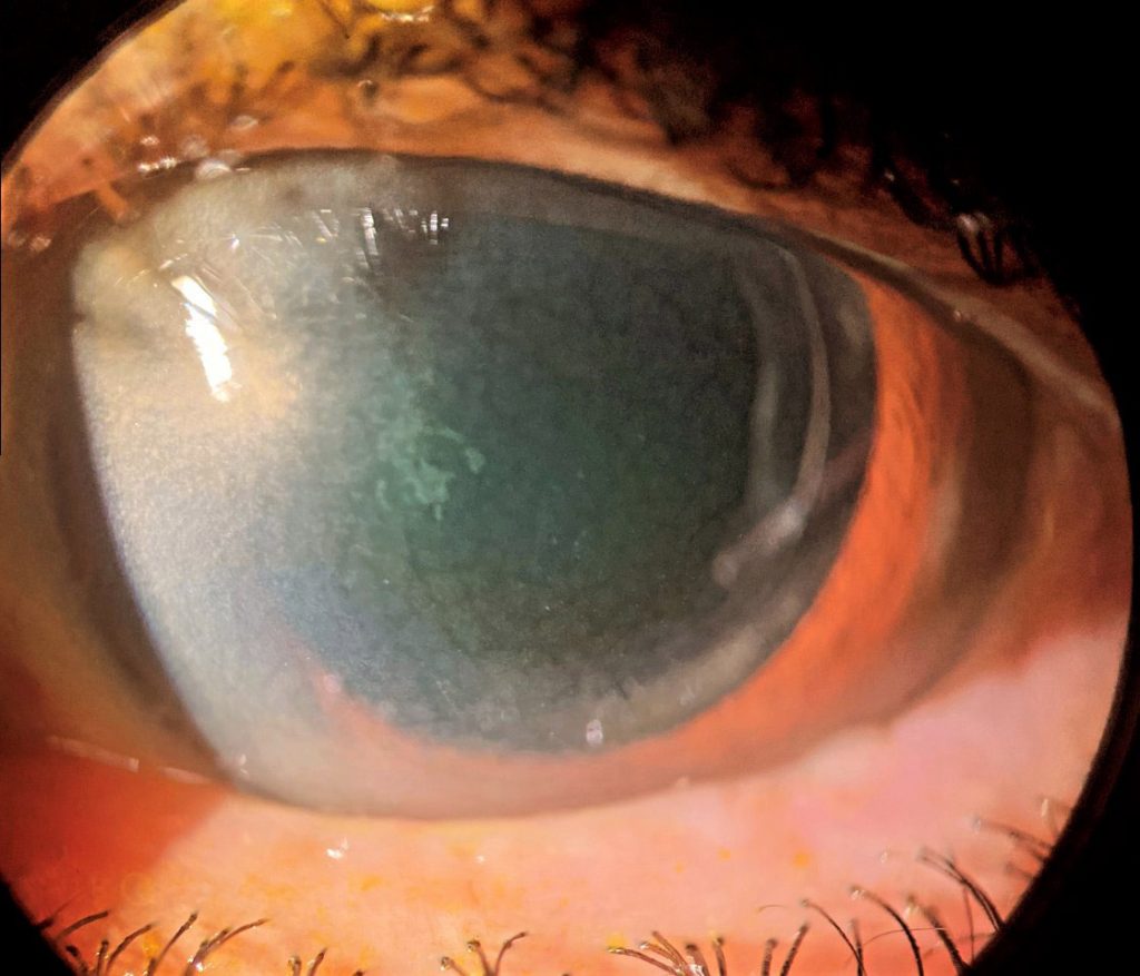
The diagnosis is made at the slit lamp.
- Polygonal grey-white opacities in the central stroma
- Clear intervening spaces creating a mosaic pattern
- Best appreciated with broad oblique illumination and retroillumination
The finding should be documented, but on its own it does not explain reduced acuity.
Associations
Crocodile shagreen is most strongly associated with advancing age.
Additional reported associations include:
- Band keratopathy
- Previous traumatic corneal injury
- Pseudoxanthoma elasticum (isolated reports)
- Glaucoma (isolated reports)
- Polymorphic amyloid degeneration (isolated reports)
These associations are largely incidental and do not change the benign course of the finding.
Differential Diagnosis
Several corneal conditions can produce a similar deep stromal pattern:
- Central cloudy dystrophy of François — clinically almost indistinguishable from posterior crocodile shagreen, but inherited (autosomal dominant) rather than age-related
- Pre-Descemet corneal dystrophy
- Cornea farinata
- Band keratopathy (for the anterior form)
- Posterior polymorphous corneal dystrophy
Because these entities differ in inheritance and prognosis, clinical context and family history are the most useful discriminators.
Management
Crocodile shagreen requires no treatment.
- The finding does not reduce vision and does not progress meaningfully
- Reassurance is the primary management
- Any visual complaint should be investigated for a separate cause, most commonly cataract
Surgical intervention is essentially never indicated for crocodile shagreen itself.
Prognosis
The prognosis is excellent.
- The condition is benign and visually insignificant
- It remains stable or progresses only very slowly over years
- Recognizing it prevents unnecessary imaging, referral, and patient anxiety
In practice, the correct response to crocodile shagreen is to document it, exclude other causes of any symptoms, and reassure the patient.
Would you like to capture slit-lamp findings like crocodile shagreen with your smartphone?


Document what you see
Two smartphone imaging tools built for everyday clinic use — one for the slit lamp, one for the fundus.
From Choroida — the team behind this siteReferences
- American Academy of Ophthalmology. “Crocodile Shagreen.” EyeWiki.
- Belliveau MJ, Brownstein S, Agapitos P, Font RL. “Ultrastructural Features of Posterior Crocodile Shagreen of the Cornea.” Survey of Ophthalmology. 2009;54(5):569–575.
- Tripathi RC, Bron AJ. “Secondary anterior crocodile shagreen of Vogt.” British Journal of Ophthalmology. 1975;59(1):59.
- Krachmer JH, Mannis MJ, Holland EJ. Cornea. 2nd ed. 2005; Vol 1, p.999.
Test yourself
A few questions straight from this article.
-
How does crocodile shagreen appear at the slit lamp?
Crocodile shagreen is a mosaic of greyish-white polygonal stromal opacities with indistinct margins, separated by clearer intervening zones, resembling crocodile hide. -
What is the typical distribution of crocodile shagreen?
The finding is usually bilateral, broadly symmetric and central or paracentral, and is most often discovered incidentally on routine examination. -
Anterior crocodile shagreen is also known by which eponym?
The anterior form, known as mosaic degeneration of Vogt, involves the level of Bowman layer and the anterior stroma. -
At which corneal depth does posterior crocodile shagreen lie?
The posterior form sits in the deep posterior stroma just anterior to Descemet membrane, and is the type most often photographed and reported. -
What is thought to produce the grey opacities of crocodile shagreen?
Ultrastructural studies describe alternating normally and abnormally oriented collagen fibrils in a periodic mosaic, with light scattering at the abnormal zones creating the visible opacities. -
Which factor is crocodile shagreen most strongly associated with?
Crocodile shagreen is most strongly associated with advancing age; band keratopathy, previous trauma, pseudoxanthoma elasticum, glaucoma and polymorphic amyloid degeneration are reported associations. -
Which condition is clinically almost indistinguishable from posterior crocodile shagreen?
Central cloudy dystrophy of Francois looks almost identical but is inherited in an autosomal dominant fashion rather than being age-related, so family history is the key discriminator. -
How is crocodile shagreen best examined at the slit lamp?
The mosaic pattern of polygonal opacities with clear intervening spaces is best appreciated using broad oblique illumination and retroillumination. -
A patient with crocodile shagreen complains of blurred vision. What should the clinician do?
Crocodile shagreen does not itself reduce vision, so any visual complaint should be investigated for a separate cause, most commonly cataract. -
What is the prognosis of crocodile shagreen?
The prognosis is excellent: the condition is benign, needs no treatment, and recognising it prevents unnecessary imaging, referral and patient anxiety.

Writes and reviews the clinical articles on Educate. Every post is checked against peer-reviewed sources before it goes live.


