
CASE REPORT
Case of Cherry red spot 1 year and 7 months aged male came to the hospital brought by his mother with seizures, cough, and fever for about one week. He had gone to the hospital twice before due to otitis and pneumonia.

His parents were healthy. When the baby was one month old, it was observed that the baby had little power for holding its head or moving its limbs.
Since then, the weakness has become progressively evident. The mother noticed that the baby did not see like other children of the same age.
In the hospital initial exam, the child had a fever, rhonchi on chest auscultation, and marasmus-like secondary protein-energy malnutrition.
The neurological examination revealed psychomotor retardation, horizontal and bilateral nystagmus, muscle weakness, generalized hyperreflexia, clumsiness, and the presence of Babinski and Moro signal. Diagnosis of Tay-Sachs disease was done.
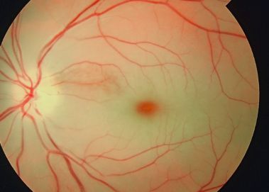
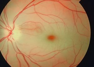
The fundoscopic examination was very difficult due to the nystagmus and showed chalk-white macular areas with a “Cherry red spot” in the center of both eyes.
DISEASE of Cherry red spot
This is a significant fundoscopic finding in the macula, observed in central retinal artery occlusion (CRAO) and a variety of lipid storage disorders.
The term refers to the appearance of a red-tinted region at the center of the macula surrounded by retinal opacification, and it consists of a clinical sign usually present within the context of thickening and loss of transparency in the posterior pole of the retina.
The appearance of Cherry red spot can result from retinal edema most commonly due to central retinal artery occlusion or traumatic retinal ischemia, which causes the perimacular tissue of the retina to appear translucent, while the fovea maintains its normal color.
In the case of lipid storage diseases, the lipids are stored in the ganglion cell layer of the retina, giving the retina a white appearance. As ganglion cells are absent at the foveola, this area retains relative transparency and contrasts with the surrounding retina.
The Cherry red spot is an important fundoscopic sign, and when found with key clinical features and a good history, often guides one to the diagnosis of the disease.
MANAGEMENT of Cherry red spot
The general management of a cherry-red spot sign is determined by the underlying pathological condition that causes the appearance of the fundoscopic finding.
In the acute setting of CRAO, treatment options are directed at resolving the CRAO and circulation, and thus maximizing visual outcome.
For various storage diseases, the management requires a team of various specialties. Generalized pearls for the management of such patients include avoiding/reducing intake of specific molecules that lead to accumulation within ganglion cells, enzyme replacement therapy, and symptomatic management.
would you have the interest to take retinal photography by smartphone?
Fundus photography is superior to fundus analysis as it enables intraocular pathologies to be photo captured and encrypted information to be shared with colleagues and patients.
Recent technologies allow smartphone-based attachments and integrated lens adaptors to transform the smartphone into a portable fundus camera.

Read more: Branch Retinal Artery Occlusion, Retinitis Pigmentosa, Bietti Crystalline Dystrophy, Arcus Senilis.
REFERENCES
- Suvarna J C, Hajela S A. Cherry-red spot. J Postgrad Med 2008;54:54-7
- Tripathy K, Patel BC. Cherry Red Spot. [Updated 2021 Feb 14]. In: StatPearls [Internet]. Treasure Island (FL): StatPearls Publishing; 2021 Jan-. Available from: https://www.ncbi.nlm.nih.gov/books/NBK539841/
- General Practice Notebook
- USMLE First AID 2010 page 417
- Leavitt, J. A., & Kotagal, S. (2007). The “Cherry Red” Spot. Pediatric Neurology, 37(1), 74-75 doi:10.1016/j.pediatrneurol.2007.04.011.
- Leavitt, J. A., & Kotagal, S. (2007). The “Cherry Red” Spot. Pediatric Neurology, 37(1), 74–75. doi:10.1016/j.pediatrneurol.2007.04.011
- Kaufman EJ, Mahabadi N, Patel BC. Hollenhorst Plaque. [Updated 2021 Feb 25]. In: StatPearls [Internet]. Treasure Island (FL): StatPearls Publishing; 2021 Jan-. Available from: https://www.ncbi.nlm.nih.gov/books/NBK470445/
- Varma DD, Cugati S, Lee AW, Chen CS. A review of central retinal artery occlusion: clinical presentation and management. Eye (Lond). 2013 Jun;27(6):688-97. doi: 10.1038/eye.2013.25. Epub 2013 Mar 8. PMID: 23470793; PMCID: PMC3682348.
