
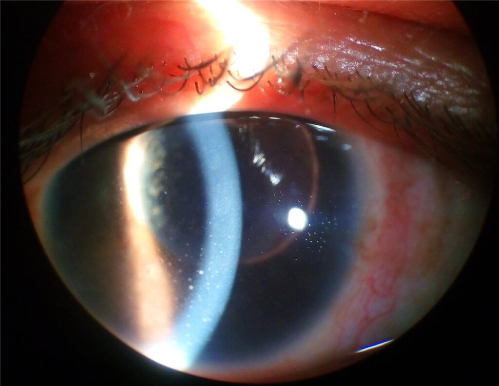
Ocular sarcoidosis is inflammation of the eye and adnexa related to systemic sarcoidosis, a multisystem granulomatous disease of unknown cause, and the eye is involved in a substantial proportion of patients with the systemic disease at some point in its course.
Sarcoidosis is genuinely called the great mimicker in ophthalmology for good reason: it can present as almost any pattern of intraocular or adnexal inflammation, which is why it belongs on the differential for uveitis that does not fit a more obvious cause.
Patterns of Ocular Involvement
Anterior uveitis is the most common ocular manifestation, and it can be either acute or, more characteristically for sarcoidosis, chronic and granulomatous, with large mutton-fat keratic precipitates on the corneal endothelium.
Posterior segment involvement includes intermediate uveitis with vitreous snowballs, retinal periphlebitis producing the classic “candlewax drippings” appearance along retinal veins, chorioretinal granulomas, and optic disc involvement, either from granuloma or from associated papilloedema in neurosarcoidosis.
Adnexal involvement includes lacrimal gland enlargement, sometimes bilateral and occasionally the presenting sign of the disease, conjunctival granulomas, and orbital involvement, which is less common but recognised.
Clinical Presentation
Symptoms depend on which part of the eye is involved. Anterior uveitis typically causes redness, pain, and photophobia, while posterior involvement more often presents with floaters and blurred vision, sometimes without significant pain or redness at all.
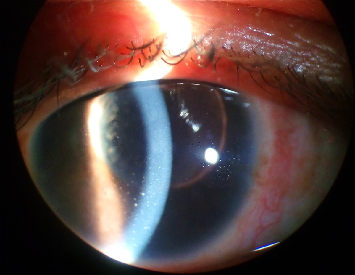
On examination, keratic precipitates on the corneal endothelium are a key finding in anterior involvement, and their size and distribution, classically large and greasy-appearing mutton-fat precipitates in granulomatous disease, is a useful diagnostic clue, though smaller, fine precipitates can also be seen and do not exclude the diagnosis.
A chronic, indolent course, bilateral involvement, and posterior synechiae from longstanding inflammation are all more suggestive of a granulomatous process like sarcoidosis than of a typical acute anterior uveitis.
Systemic symptoms, including cough, dyspnoea, skin lesions such as erythema nodosum, and lymphadenopathy, should be asked about specifically, since their presence supports the diagnosis and points toward the need for a broader systemic workup.
All-fit Slit-Lamp Adapter
Record and share exactly what you see at the slit lamp. One adapter fits any slit lamp or surgical microscope — and any smartphone.
From Choroida — the team behind this siteDiagnostic Evaluation
Chest imaging, historically chest X-ray but increasingly CT, is used to look for bilateral hilar lymphadenopathy, the classic and highly suggestive systemic finding in sarcoidosis.
Serum angiotensin-converting enzyme and serum lysozyme can support the diagnosis when elevated, though neither is fully sensitive or specific on its own, and normal levels do not exclude sarcoidosis.
Tissue biopsy showing non-caseating granulomas remains the definitive diagnostic standard, obtained from the most accessible affected site, which can include a conjunctival nodule, enlarged lacrimal gland, mediastinal lymph node, or another accessible lesion depending on where disease is evident.
Where systemic evaluation is inconclusive but clinical suspicion remains high, published diagnostic criteria for presumed ocular sarcoidosis, incorporating a combination of clinical, laboratory, and imaging features, can support a working diagnosis without tissue confirmation in selected cases.
Differential Diagnosis
- Tuberculosis, which can produce a very similar granulomatous uveitis and needs to be excluded, particularly given the opposite immunosuppressive implications of the two diagnoses
- Vogt-Koyanagi-Harada disease, another granulomatous panuveitis, distinguished by its own characteristic systemic and imaging features
- Sympathetic ophthalmia, relevant when there is a history of penetrating ocular trauma or surgery in the fellow eye
- Other causes of intermediate uveitis with vitreous snowballs, including pars planitis, where systemic sarcoidosis workup is negative
- Lymphoma, particularly when lacrimal gland or orbital involvement is prominent, which can mimic sarcoid adnexal disease and sometimes requires biopsy to distinguish
Management
Topical corticosteroids and cycloplegics are first-line for anterior uveitis, following standard principles of uveitis management, with the intensity and duration guided by the severity of inflammation.
Periocular or intravitreal corticosteroid injections are used for posterior segment inflammation not adequately controlled with topical treatment alone.
Systemic corticosteroids are used for significant posterior or bilateral disease, and for managing extraocular systemic manifestations, coordinated with the physician managing the patient’s systemic sarcoidosis.
Steroid-sparing immunosuppressive agents, including methotrexate and other conventional agents, and biologic therapy in refractory cases, are used for chronic or recurrent disease requiring long-term control, again in coordination with the patient’s broader medical team rather than managed by ophthalmology in isolation.
Prognosis
Visual outcome is generally good with appropriate treatment for isolated anterior uveitis, but posterior segment involvement, particularly with macular oedema, retinal vasculitis, or optic nerve involvement, carries a greater risk of permanent visual loss if not controlled promptly.
Chronic or recurrent disease is common, and many patients need long-term surveillance and, in some cases, ongoing immunosuppression to prevent relapse.
Because sarcoidosis is a systemic disease, ocular prognosis is only part of the picture. Coordinated care between ophthalmology and the physicians managing systemic disease, particularly pulmonary and, where relevant, neurological involvement, is central to the overall outcome for the patient.


Document what you see
Two smartphone imaging tools built for everyday clinic use — one for the slit lamp, one for the fundus.
From Choroida — the team behind this siteReferences
- Herbort CP, Rao NA, Mochizuki M. International criteria for the diagnosis of ocular sarcoidosis: results of the first International Workshop on Ocular Sarcoidosis. Ocular Immunology and Inflammation. 2009.
- Pasadhika S, Rosenbaum JT. Ocular sarcoidosis. Clinics in Chest Medicine. 2015.
- Jamilloux Y, Kodjikian L, Broussolle C, Seve P. Sarcoidosis and uveitis. Autoimmunity Reviews. 2014.
- Ocular Sarcoidosis. EyeWiki, American Academy of Ophthalmology.
- Sarcoidosis. StatPearls, NCBI Bookshelf.
Test yourself
A few questions straight from this article.
-
Which is the most common ocular manifestation of sarcoidosis?
Anterior uveitis is the commonest ocular manifestation, and is characteristically chronic and granulomatous in sarcoidosis rather than acute. -
Which corneal endothelial finding is classic for granulomatous uveitis in ocular sarcoidosis?
Large, greasy mutton-fat keratic precipitates typify granulomatous disease, although finer precipitates can also occur and do not exclude sarcoidosis. -
Retinal periphlebitis in ocular sarcoidosis classically produces which fundus appearance?
Sarcoid retinal periphlebitis produces the classic candlewax drippings appearance of exudate distributed along the retinal veins. -
Which adnexal finding may be the presenting sign of ocular sarcoidosis?
Lacrimal gland enlargement, which may be bilateral, is a recognised adnexal manifestation and is occasionally how the disease first declares itself. -
Chest imaging in suspected ocular sarcoidosis is performed to look for which classic finding?
Chest X-ray or, increasingly, CT is used to detect bilateral hilar lymphadenopathy, the classic and highly suggestive systemic finding. -
How should a normal serum angiotensin-converting enzyme level be interpreted in suspected ocular sarcoidosis?
Serum ACE and lysozyme support the diagnosis when raised, but neither is fully sensitive or specific, so normal values do not exclude the disease. -
What remains the definitive diagnostic standard for sarcoidosis?
Biopsy demonstrating non-caseating granulomas, taken from the most accessible affected site, is the definitive standard for diagnosis. -
Which mimic of ocular sarcoidosis must be excluded because it carries opposite immunosuppressive implications?
Tuberculosis can produce a very similar granulomatous uveitis, and the two diagnoses carry opposite implications for immunosuppressive treatment. -
What is first-line treatment for sarcoid anterior uveitis?
Topical steroids plus cycloplegics are first-line, with intensity and duration set by the severity of the inflammation. -
Which pattern of ocular sarcoidosis carries the greatest risk of permanent visual loss?
Isolated anterior uveitis generally does well, whereas macular oedema, retinal vasculitis or optic nerve involvement risks permanent loss unless controlled promptly.

Writes and reviews the clinical articles on Educate. Every post is checked against peer-reviewed sources before it goes live.


