
CASE REPORT
A 45-year-old male software engineer with no significant ocular history or known systemic diseases presented to an ophthalmology clinic complaining of sudden-onset, severe pain, redness, and decreased vision in his right eye.
The symptoms had initiated two days prior and had progressively worsened. Clinical examination revealed visual acuity of counting fingers at 1 meter in the right eye and 20/20 in the left eye.
Anterior segment examination of the right eye showed conjunctival injection, corneal edema, and a 2+ anterior chamber cell, while the left eye appeared unremarkable.
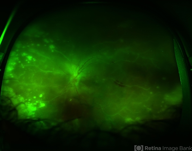
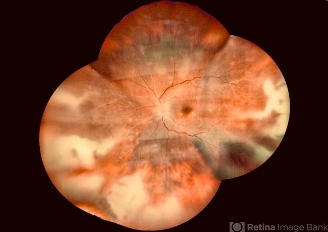
Fundus examination of the right eye indicated severe vitritis, retinal necrosis involving the peripheral retina, and multiple areas of retinal hemorrhages with a swollen optic disc.
Fluorescein angiography of the right eye demonstrated delayed arteriovenous transit time, areas of capillary non-perfusion, and leakage from the optic disc, while the left eye had a normal angiogram.
Polymerase Chain Reaction (PCR) testing confirmed the presence of Varicella-Zoster Virus (VZV) DNA in the aqueous humor of the affected eye, leading to a diagnosis of Acute Retinal Necrosis (ARN) in the right eye.
DISEASE
Acute retinal necrosis (ARN) is an inflammatory condition that may present as panuveitis. The principal causative viral agents have been found to be Varicella Zoster Virus (VZV) as well as Herpes Simplex Virus (HSV-1 and HSV-2) via polymerase chain reaction (PCR) testing of intraocular fluid.

In 1971, Urayama and colleagues at Tohoku University in Japan reported 6 cases of a syndrome characterized by unilateral panuveitis, vitritis, acute necrotizing retinitis, retinal arteritis, and rhegmatogenous retinal detachment.
Young and Bird proposed the name of bilateral acute retinal necrosis. In Japan, the name ‘Kirisawa-type uveitis’ had been used for Acute retinal necrosis (ARN).
ARN is believed to be caused by a viral infection. Most often the causative viral agent is either VZV or less commonly HSV.
Older patients more often present with VZV or HSV-1 infections while younger patients more often present with HSV-2 infections.
Acute retinal necrosis (ARN) has also been reported in case reports to be associated with cytomegalovirus (CMV) and Epstein-Barr virus (EBV), but there is limited evidence to suggest these viruses cause ARN.
This clinical entity is uncommon in the pediatric population. Most often it affects healthy, immunocompetent adults in their fifth to seventh decade. A clear racial or gender predilection has not been consistently demonstrated.
Fundus Explorer Pro
Photograph the retinal findings described here with the phone already in your pocket — 22 D optics and built-in illumination in one handheld unit.
From Choroida — the team behind this siteMANAGEMENT
General treatment
Without intervention, the active phase of the disease can last between 6-12 weeks, while treatment with antivirals and/or steroids may reduce the latency to 4-6 weeks.

Treatment of Acute retinal necrosis (ARN) is complex and should be individualized based on the overall clinical picture and vitreoretinal findings.
Prospective, randomized controlled trials have not been performed given the rarity of the disease; thus, treatment recommendations are based on case reports/series.
Treatment modalities may include antiviral, anti-inflammatory, and antithrombotic therapy in cases with ischemic optic neuropathy, as well as laser photocoagulation for retinal detachment prophylaxis or surgery for retinal detachment repair.
Medical therapy
Antiviral therapy for Acute retinal necrosis (ARN) can be administered either systemically (intravenously or orally) or via intravitreal injection.
While intravenous administration of agents like acyclovir was common in the past, recent approaches favor the use of oral antivirals such as famciclovir, valacyclovir, or valganciclovir as initial induction agents.
Monitoring renal function is essential during systemic antiviral treatment. Intravitreal delivery of ganciclovir or foscarnet is a common practice and may have greater therapeutic efficacy when combined with systemic antiviral therapy.
The use of steroids and antithrombotic therapy is controversial and should be decided at the treating physician’s discretion, with caution about initiating steroids alone during the acute retinitis stage.
Commonly used systemic antiviral agents include acyclovir, famciclovir, valacyclovir, ganciclovir, and valganciclovir. Intravitreal administration of ganciclovir and foscarnet can be beneficial in cases threatening the macula or optic disc.
Steroids, such as prednisone, may be considered after the initiation of antiviral therapy, and aspirin could minimize vascular thrombosis.
Topical steroids and cycloplegics may be beneficial depending on anterior segment inflammation, while topical antiviral therapy has shown inefficacy in ARN treatment.
Medical follow-up
Patients with unilateral Acute retinal necrosis (ARN) should be closely followed with a dilated examination of both of their eyes. Approximately 50-75% of patients with ARN will go on to develop a retinal detachment.
Reports of second eye involvement range from 3-35%, with variable involvement attributable to etiologic agents, course of therapy, and immune status.
The antiviral therapy reduces the duration of active disease and reduces the chance of involvement of the fellow eye. However, the retinal detachment may not be reduced by antiviral therapy.
Surgery
The role of prophylactic laser retinopexy or early PPV is unknown at this time. Some retina specialists advocate the use of prophylactic lasers along the posterior margin of the junction of necrotic and non-necrotic retina to prevent the incidence of retinal detachment.
The rates of success in preventing retinal detachment are variable, and consensus does not exist on whether laser demarcation should be employed.
This may be due to the confounding fact that eyes in which laser can be successfully performed typically have optically clearer vitreous, less inflammation, and thereby less risk for developing retinal detachment.
Vitrectomy with or without the use of a scleral buckle, with either gas or silicone oil tamponade, is usually reserved for those cases in which traction-rhegmatogenous retinal detachment occurs.


Document what you see
Two smartphone imaging tools built for everyday clinic use — one for the slit lamp, one for the fundus.
From Choroida — the team behind this siteREFERENCES
- Urayama A, Yamada N, Sasaki T, et al. Unilateral acute uveitis with retinal periarteritis and detachment. Jpn J Clin Ophthalmol 1971;25:607–19.
- Young NJ, Bird AC. Bilateral acute retinal necrosis. Br J Ophthalmol 1978;62(9):581–90.
- Tripathy K, Sharma YR, Gogia V, Venkatesh P, Singh SK, Vohra R. Serial ultra wide field imaging for following up acute retinal necrosis cases. Oman J Ophthalmol. 2015;8:71-2.
- Chawla R, Tripathy K, Sharma YR, Venkatesh P, Vohra R. Periarterial Plaques (Kyrieleis’ Arteriolitis) in a Case of Bilateral Acute Retinal Necrosis. Semin Ophthalmol. 2017;32 :251-252.
- Tripathy K, Chawla R, Venkatesh P, Sharma YR, Vohra R. Ultrawide Field Imaging in Uveitic Non-dilating Pupils. J Ophthalmic Vis Res. 2017 ;12:232-233.
- Schoenberger SD, Kim SJ, Thorne JE, Mruthyunjaya P, Yeh S, Bakri SJ, Ehlers JP. Diagnosis and Treatment of Acute Retinal Necrosis: A Report by the American Academy of Ophthalmology. Ophthalmology. 2017 Mar;124(3):382-392.
- Risseeuw S, de Boer JH, Ten Dam-van Loon NH, van Leeuwen R. Risk of Rhegmatogenous Retinal Detachment in Acute Retinal Necrosis With and Without Prophylactic Intervention. Am J Ophthalmol. 2019 Oct;206:140-148.
