
Case report
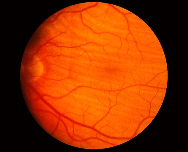
Case of Choroidal Folds 69-year-old female patient presented with a recent-onset slight decrease in visual acuity in her left eye. Her past ocular history was clear. Regarding her medical history, she had only hypertension, treated with per os medications.

The patient underwent a complete ophthalmological examination. The best-corrected visual acuity was 6/6 in the right eye and 6/7.5 in the left eye. Intraocular pressure was 16 mm Ηg in both eyes.
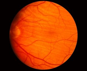
On fundoscopy, chorioretinal folds were noticed.
Disease of Choroidal Folds
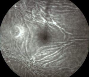
These are lines, grooves, or striae predominantly involving the posterior pole of the eye which appear as alternating light and dark lines on fluorescein angiography.
They are often arranged in a parallel and horizontal fashion but maybe vertical, oblique, or irregular.
These folds tend to vary in length and width, rarely extending beyond the equator and may extend into the neurosensory retina, in which case they can be called chorioretinal folds.
Management
There is sufficient evidence that as soon as choroidal folds are recognized, a prompt systemic investigation for a specific diagnosis or cause is mandatory in order to diagnose treatable causes or to exclude potentially serious conditions.
would you have the interest to take Retinal imaging by smartphone ?
Fundus photography is superior to fundus analysis as it enables intraocular pathologies to be photo captured and encrypted information to be shared with colleagues and patients.
Recent technologies allow smartphone-based attachments and integrated lens adaptors to transform the smartphone into a portable fundus camera help in diagnose Choroidal Folds and others easily.
Read about Choroidal Osteoma, Birdshot Retinochoroidopathy, Optociliary Shunt Vessels, Morgagnian Cataract.

References
- Bowling B. Kanskiʼs Clinical Ophthalmology: A Systematic Approach. Eighth Edition. Cornea. 2016.
- Bullock JD, Egbert PR. The origin of choroidal folds a clinical, histopathological, and experimental study. Doc Ophthalmol. 1974. doi:10.1007/BF00147262
- Cangemi FE, Trempe CL, Walsh JB. Choroidal folds. Am J Ophthalmol. 1978. doi:10.1016/0002-9394(78)90243-X
- Dailey RA, Mills RP, Stimac GK, Shults WT, Kalina RE. The Natural History and CT Appearance of Acquired Hyperopia with Choroidal Folds. Ophthalmology. 1986. doi:10.1016/S0161-6420(86)33577-2
