
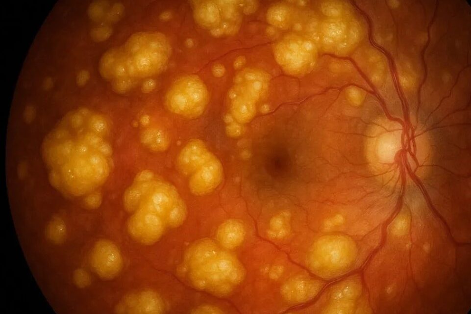
Ocular tuberculosis is a genuine diagnostic challenge because it can mimic nearly every other uveitis entity.
It frequently occurs — especially outside high-burden countries — without any obvious active pulmonary disease, making it easy to overlook unless specifically considered.
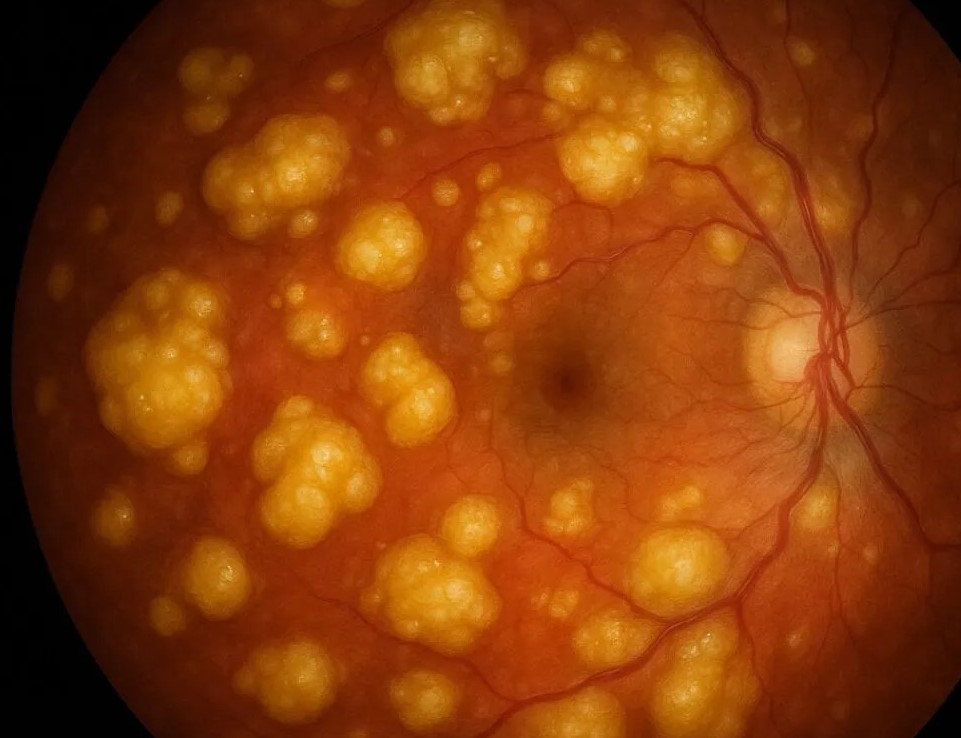
It arises either from direct hematogenous spread of Mycobacterium tuberculosis to ocular tissue or from an immune hypersensitivity reaction to mycobacterial antigens without the organism itself being present in the eye, and distinguishing these two mechanisms has real implications for how confidently a clinician can rely on the response to treatment as diagnostic confirmation.
Clinical Patterns
- Choroidal tubercles — small, yellow-white, round choroidal lesions, often multiple, seen in disseminated (miliary) TB; a classic finding worth specifically searching for on dilated exam in any patient being evaluated for suspected miliary tuberculosis
- Tuberculoma — a larger, solitary choroidal or, less commonly, retinal mass lesion that can mimic a tumor
- Serpiginous-like choroiditis — a multifocal, geographic pattern of choroiditis that can closely resemble idiopathic serpiginous choroidopathy, one of the more important mimics to distinguish given very different treatment implications
- Granulomatous anterior uveitis — mutton-fat keratic precipitates and iris nodules, similar in appearance to sarcoidosis and other granulomatous causes
- Retinal vasculitis — periphlebitis, sometimes associated with vitreous hemorrhage, historically described as Eales disease in some populations before TB was more directly implicated
- Phlyctenular keratoconjunctivitis — a hypersensitivity reaction to mycobacterial antigen, discussed in more detail in its own dedicated article on this site
This range of possible presentations is precisely why ocular TB has earned a reputation as a “great masquerader” alongside conditions like sarcoidosis and syphilis, and why it deserves active consideration in essentially any atypical, treatment-resistant, or recurrent uveitis, particularly in patients from or with relevant travel history to higher-burden regions, or with known immunosuppression that increases susceptibility to reactivation of latent infection.
Why the Diagnosis Is Difficult
No single test reliably confirms active intraocular tuberculosis, because direct isolation of the organism from ocular tissue or fluid is difficult and often unsuccessful even when the diagnosis is ultimately correct.
Standard tests (tuberculin skin test, interferon-gamma release assay) indicate prior exposure or latent infection rather than proving the eye disease in front of the clinician is actually caused by TB rather than another process in a patient who happens to have been exposed to the organism at some point.
This is why ocular TB is frequently a diagnosis reached through a combination of a compatible clinical picture, supportive but non-confirmatory testing, and, often, a therapeutic trial.
Fundus Explorer Pro
Photograph the retinal findings described here with the phone already in your pocket — 22 D optics and built-in illumination in one handheld unit.
From Choroida — the team behind this siteDiagnostic Evaluation
- Tuberculin skin test or interferon-gamma release assay (QuantiFERON-TB or similar) to establish exposure/latent infection
- Chest imaging to look for evidence of pulmonary disease, active or old, though a normal chest X-ray does not exclude ocular TB
- PCR testing of aqueous or vitreous fluid for mycobacterial DNA where available, which can be supportive when positive but has limited sensitivity, so a negative result does not exclude the diagnosis
- Systematic exclusion of other granulomatous uveitis causes — sarcoidosis, syphilis, and other infectious and autoimmune entities that can present similarly
Because so much of the workup consists of tests that are supportive rather than definitively confirmatory, building a complete picture from the clinical pattern, exposure history, and every available test result together — rather than searching for a single decisive positive result — is the realistic approach in most suspected cases.
International Consensus Approach
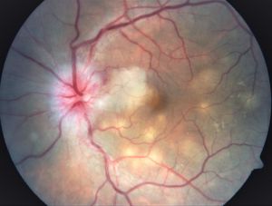
Given the diagnostic difficulty, expert consensus groups have proposed structured criteria combining the clinical pattern — choroidal tubercles and serpiginous-like choroiditis being considered the most specific findings, with the other patterns above weighted accordingly — with supportive systemic evidence (positive TST/IGRA, chest imaging findings) to categorize cases as confirmed, probable, or possible ocular tuberculosis.
This is because a purely binary “TB or not TB” framework does not fit the reality of how these cases actually present.
Management
Standard multidrug antituberculous therapy (typically a four-drug regimen — isoniazid, rifampin, ethambutol, pyrazinamide — for an initial phase, followed by a longer continuation phase) is the mainstay of treatment for confirmed or strongly probable ocular TB, generally coordinated with infectious disease or pulmonology given the systemic nature of the underlying infection and the need for treatment monitoring, including liver function and visual toxicity screening (ethambutol carries its own, separate risk of optic neuropathy that needs to be distinguished from the disease itself).
Corticosteroids are frequently added to control the ocular inflammatory response, but only after antituberculous therapy has been started or is being given concurrently, because steroids alone in unrecognized active TB risk worsening systemic disease.
A favorable response to antituberculous therapy — resolution of the ocular inflammation — is itself often used as supportive, retrospective evidence for the diagnosis in cases where the initial workup was inconclusive.
Given how heavily ethambutol-related optic neuropathy relies on clinical monitoring for early detection, baseline and periodic visual acuity and color vision testing are a routine part of care throughout treatment, so that a genuinely reversible toxic optic neuropathy is caught and the drug adjusted before permanent visual loss develops, and so that any new visual change during treatment is not mistakenly attributed to the underlying ocular TB when it is in fact a medication side effect requiring its own separate management.


Document what you see
Two smartphone imaging tools built for everyday clinic use — one for the slit lamp, one for the fundus.
From Choroida — the team behind this siteReferences
- Gupta V, Gupta A, Rao NA. Intraocular tuberculosis — an update. Survey of Ophthalmology.
- Agrawal R, Gupta B, Gonzalez-Lopez JJ, et al. The role of anti-tubercular therapy in patients with presumed ocular tuberculosis. Ocular Immunology and Inflammation.
- Collaborative Ocular Tuberculosis Study (COTS) consensus reports on ocular tuberculosis nomenclature and diagnostic criteria.
- American Academy of Ophthalmology. Basic and Clinical Science Course, Section 9: Intraocular Inflammation and Uveitis.
Test yourself
A few questions straight from this article.
-
By which routes does ocular tuberculosis arise?
The eye may be seeded directly by Mycobacterium tuberculosis, or inflamed by an immune hypersensitivity reaction to mycobacterial antigen with no organism present in the eye. -
What is the classic choroidal finding in disseminated miliary tuberculosis?
Choroidal tubercles are small round yellow-white lesions, frequently multiple, and are worth searching for on dilated examination in any patient assessed for miliary tuberculosis. -
Which entity does tubercular serpiginous-like choroiditis most closely resemble?
The multifocal geographic choroiditis of ocular TB closely mimics idiopathic serpiginous choroidopathy, an important distinction because the treatment implications are very different. -
Which anterior segment picture can ocular tuberculosis produce?
Ocular TB can cause a granulomatous anterior uveitis with mutton-fat keratic precipitates and iris nodules, an appearance shared with sarcoidosis and other granulomatous causes. -
What does a positive interferon-gamma release assay establish in suspected ocular tuberculosis?
TST and IGRA show exposure or latent infection only; they cannot prove that the uveitis in front of you is tuberculous rather than another process in a previously exposed patient. -
How should a negative aqueous PCR for mycobacterial DNA be interpreted?
PCR of aqueous or vitreous is supportive when positive but has limited sensitivity, so a negative result leaves the diagnosis open, just as a normal chest X-ray does. -
How do consensus criteria categorize suspected ocular tuberculosis?
Expert groups combine the clinical pattern with supportive systemic evidence to grade cases as confirmed, probable or possible, since a binary TB-or-not framework does not fit these presentations. -
What is the mainstay of treatment for confirmed or strongly probable ocular tuberculosis?
Isoniazid, rifampin, ethambutol and pyrazinamide are given for an initial phase followed by a longer continuation phase, usually coordinated with infectious disease or pulmonology. -
When may corticosteroids be added for the inflammation of ocular tuberculosis?
Steroids control the ocular inflammatory response, but giving them alone in unrecognized active tuberculosis risks worsening the systemic infection. -
Which monitoring detects ethambutol optic neuropathy during treatment of ocular tuberculosis?
Ethambutol carries its own risk of optic neuropathy, so acuity and color vision are checked throughout treatment to catch a reversible toxicity before permanent loss occurs.
