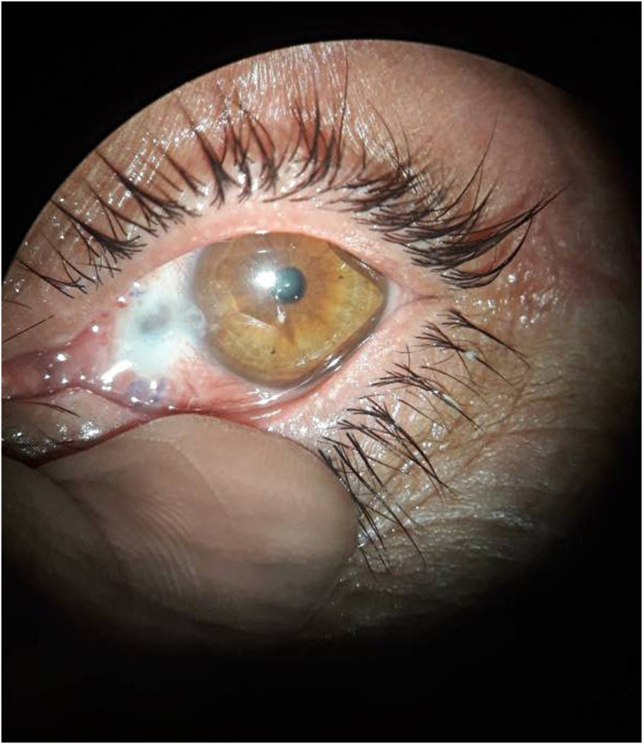
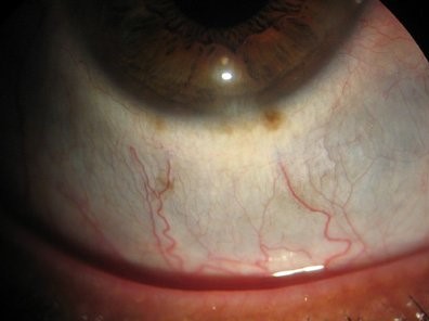
Scleral thinning is a loss of tissue in the wall of the eye, and it appears as a blue-gray area where the dark uvea shows through the sclera.
It can be harmless, as in a stable area of old inflammation, or dangerous, when it heralds a necrotizing process that can lead to perforation.
Also, the task is to separate those groups, and to find the cause.
What Is the Sclera?
The sclera is a dense, avascular connective tissue made mainly of collagen, with some elastic fibers and few cells.
It protects the eye and maintains its shape, and it is also covered by the episclera and the conjunctiva.
In addition, the sclera varies in thickness, with the thinnest areas at the insertion of the rectus muscles and near the equator (see scleritis).
When tissue is lost, the underlying uvea becomes visible, and the eye wall weakens.
Causes of Scleral Thinning
- Inflammatory: necrotizing scleritis, scleromalacia perforans, and posterior scleritis (see necrotizing scleritis, scleromalacia perforans, and posterior scleritis)
- Systemic autoimmune disease: rheumatoid arthritis, granulomatosis with polyangiitis, relapsing polychondritis, and lupus (see granulomatosis with polyangiitis)
- Infectious: herpes zoster, tuberculosis, syphilis, fungal, and bacterial scleritis
- Surgical and iatrogenic: after pterygium surgery with mitomycin C or radiation, strabismus surgery, scleral buckle, and glaucoma drainage devices
- Traumatic and chemical: injuries and burns
- Congenital and genetic: osteogenesis imperfecta, Ehlers-Danlos syndrome, and Marfan syndrome (see Ehlers-Danlos syndrome)
- Degenerative: high myopia and staphyloma (see staphyloma)
- Drug-related: long-term topical steroids and some other agents
All-fit Slit-Lamp Adapter
Record and share exactly what you see at the slit lamp. One adapter fits any slit lamp or surgical microscope — and any smartphone.
From Choroida — the team behind this siteClinical Features of Scleral Thinning
- A blue-gray or dark patch of sclera, with or without redness and pain
- Pain, which is severe in necrotizing scleritis, and absent in scleromalacia perforans
- Avascular zones and vessel changes around the area
- Bulging of the uvea, forming a staphyloma, in advanced thinning
- Possible perforation, with uveal prolapse and a soft eye
An inflamed, painful thinning is an emergency, whereas painless, stable thinning may be an old process.
Evaluation for Scleral Thinning
Take a careful history of pain, previous surgery, trauma, and systemic disease.
Also, examine the sclera in daylight and at the slit lamp, and note the extent, location, and signs of inflammation.
Anterior segment OCT and ultrasound biomicroscopy measure the thickness of the sclera, and B-scan can also show posterior thinning and scleral changes (see anterior segment OCT and B-scan ultrasonography).
In addition, systemic tests depend on the picture: rheumatoid factor, anti-CCP, ANCA, antinuclear antibodies, inflammatory markers, tuberculosis and syphilis tests, and chest imaging.
Also, refer to a rheumatologist for suspected systemic disease.
Management
Treatment depends on the cause.
- Inflammatory disease: systemic corticosteroids and immunosuppressants, with cyclophosphamide, rituximab, or other agents in necrotizing scleritis
- Infection: specific antimicrobial therapy
- Postsurgical thinning: stop toxic agents, treat dry eye, and observe or patch
- Impending or actual perforation: a scleral or other patch graft, such as donor sclera, pericardium, or fascia, with coverage by conjunctiva or amniotic membrane (see amniotic membrane transplantation)
- Protection: avoid trauma, use eye shields or glasses, and avoid pressure on the area
Surgery does not control the inflammation, and the underlying disease must be treated at the same time.
Prognosis
Necrotizing scleritis has a risk of vision loss and a significant association with systemic disease, and some patients have a higher risk of mortality when vasculitis is present.
Early recognition and prompt immunosuppression improve the outcome.
Patients with stable thinning need monitoring and advice about trauma, since the eye wall is weak.
Patients should be taught the warning signs of a flare, such as increasing pain and redness.
Postsurgical Scleral Thinning
After pterygium excision with mitomycin C or beta radiation, the sclera at the surgical site can become avascular and thin, with melting that may occur weeks to years later.
Early signs include persistent redness and a necrotic base, and the risk is also higher with larger doses and longer application times.
Treatment includes lubricants, protection, and, in severe cases, a patch graft with amniotic membrane or conjunctiva (see conjunctival autograft pterygium surgery).
Scleromalacia Perforans
This is a painless thinning of the sclera that occurs in long-standing rheumatoid arthritis, usually in older women.
The sclera becomes necrotic without much inflammation, and the uvea shows through the thinned area, but perforation is rare unless the eye is injured.
Management concentrates on control of the systemic disease and protection of the eye.
Staphyloma
A staphyloma is a bulge of thinned sclera lined by uvea, and it may be anterior, intercalary, equatorial, or posterior.
Posterior staphyloma is also common in pathologic myopia (see posterior staphyloma).
The bulge reflects loss of scleral integrity, and the eye may also be at risk of further thinning.
Follow-Up
Measure the thickness and extent of the thinned area at intervals, using photographs and OCT, and look for progression.
Advise protective eyewear, avoid contact sports, and treat any inflammation without delay.
For the latest studies, see the PubMed results for scleral thinning.


Document what you see
Two smartphone imaging tools built for everyday clinic use — one for the slit lamp, one for the fundus.
From Choroida — the team behind this siteReferences
- Watson PG, Young RD. Scleral structure, organisation and disease: a review. Exp Eye Res. 2004;78:609-623.
- Sainz de la Maza M, Molina N, Gonzalez-Gonzalez LA, et al. Clinical characteristics of a large cohort of patients with scleritis and episcleritis. Ophthalmology. 2012;119:43-50.
- Salmon JF. Kanski’s Clinical Ophthalmology: A Systematic Approach. 9th ed. Elsevier; 2020.
- American Academy of Ophthalmology. Basic and Clinical Science Course, Section 9: Uveitis and Ocular Inflammation. AAO; current edition.

Writes and reviews the clinical articles on Educate. Every post is checked against peer-reviewed sources before it goes live.