
Leukemic retinopathy refers to the range of fundus changes seen in patients with acute or chronic leukemia, arising from a combination of anemia, thrombocytopenia, hyperviscosity, and direct leukemic infiltration of ocular tissue.
It is common enough in active leukemia that a dilated fundus exam is a reasonable part of the initial workup, and striking enough that recognizing the pattern can occasionally be the first clue to an undiagnosed hematologic malignancy.
Roth spots are one feature of this picture, but leukemic retinopathy is broader than that single finding and deserves its own framework.
Both acute and chronic leukemias can produce these findings, though the acute leukemias tend to present with more florid, rapidly evolving fundus changes given how quickly the underlying blood counts can deteriorate.
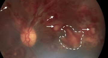
Pathophysiology
Most of the retinal findings in leukemia trace back to the same underlying hematologic derangements that define the disease: severe anemia reduces oxygen-carrying capacity, thrombocytopenia impairs normal vascular integrity, and extreme leukocytosis raises blood viscosity enough to sludge flow through the retinal microvasculature.
A smaller but clinically important subset of findings comes from direct infiltration of leukemic cells into the retina, choroid, or optic nerve, which is a genuinely different process from the hemorrhages and vascular changes driven by the systemic blood abnormalities.
Distinguishing infiltrative disease from the more common hemorrhagic picture matters, because leukemic infiltration of the optic nerve in particular is a marker of poor prognosis and often prompts urgent treatment.
Clinical Findings
- Intraretinal, preretinal, and subhyaloid hemorrhages, often more numerous and more posteriorly located than the hemorrhages seen in typical hypertensive or diabetic disease
- White-centered hemorrhages, the classic Roth spot appearance, where the pale center reflects a mix of fibrin, platelet-leukocyte aggregates, or a leukemic cell collection rather than infection specifically
- Cotton wool spots from microvascular ischemia, frequently more numerous than expected for the patient’s age or vascular risk factors
- Perivascular infiltrates and vascular sheathing, sometimes representing direct leukemic cell infiltration along the vessel wall
- Optic nerve infiltration, presenting as disc swelling with associated vision loss, distinct from papilledema and usually indicating a need for prompt orbital or systemic treatment
Fundus Explorer Pro
Photograph the retinal findings described here with the phone already in your pocket — 22 D optics and built-in illumination in one handheld unit.
From Choroida — the team behind this siteWhy the Fundus Exam Matters in Leukemia
Retinal hemorrhages and infiltrates can appear before a leukemia diagnosis is made, which occasionally puts the ophthalmologist in the position of raising the alarm for an unrecognized hematologic disease.
In a patient already known to have leukemia, new or worsening fundus findings can signal disease relapse, since active leukemic infiltration often correlates with a rising peripheral blast count or systemic recurrence after a period of remission.
Optic nerve infiltration specifically deserves urgent attention, because it behaves differently from the more indolent hemorrhagic findings and can progress to significant, sometimes irreversible, vision loss if treatment is delayed.
Differential Diagnosis
- Hypertensive retinopathy — flame hemorrhages and cotton wool spots overlap in appearance, but the systemic context and blood counts separate the two
- Diabetic retinopathy — dot-blot hemorrhages and microaneurysms follow a different distribution and typically show a much longer, more chronic vascular history
- Anemia of any other cause — the hemorrhagic picture from severe anemia alone can resemble leukemic retinopathy closely, which is exactly why the peripheral smear and full blood count are needed to tell them apart
- Central retinal vein occlusion — usually unilateral, sectoral or diffuse but confined to one venous distribution, unlike the more bilateral, diffuse pattern typical of leukemic retinopathy
- Infectious retinitis with true septic Roth spots, such as bacterial endocarditis, which shares the white-centered hemorrhage appearance but arises from a different systemic process entirely
Evaluation
The diagnosis is usually made in the context of a known or suspected hematologic workup, correlating the fundus appearance with a complete blood count, peripheral smear, and, when indicated, bone marrow biopsy.
Optical coherence tomography and fundus photography help document the extent and location of hemorrhages and infiltrates for baseline comparison and for tracking response once systemic treatment begins.
When optic nerve involvement is suspected, neuroimaging and, in some cases, cerebrospinal fluid analysis are pursued in coordination with hematology-oncology, since central nervous system leukemia and optic nerve infiltration frequently travel together.
Management
There is no separate ocular-specific treatment for most of the hemorrhagic findings in leukemic retinopathy: they improve as the underlying leukemia responds to systemic chemotherapy and as the blood counts normalize.
Optic nerve infiltration is treated more urgently, typically with local radiotherapy directed at the nerve in addition to systemic or intrathecal chemotherapy, reflecting both its distinct mechanism and its higher stakes for vision.
Ongoing ophthalmic follow-up during active treatment helps track resolution of hemorrhages and catch any new infiltrative change early, particularly around periods when systemic disease control is uncertain.
Choroidal infiltration, less common than retinal involvement, can also occur and is usually picked up incidentally on imaging obtained for the more visible retinal changes rather than as an isolated presenting complaint.


Document what you see
Two smartphone imaging tools built for everyday clinic use — one for the slit lamp, one for the fundus.
From Choroida — the team behind this siteReferences
- Reddy SC, Jackson N, Menon BS. Ocular involvement in leukemia: a study of 288 cases. Ophthalmologica.
- Schachat AP, Markowitz JA, Guyer DR, et al. Ophthalmic manifestations of leukemia. Archives of Ophthalmology.
- American Academy of Ophthalmology. Basic and Clinical Science Course, Section 12: Retina and Vitreous.
- Sharma T, Grewal J, Gupta S, Murray PI. Ophthalmic manifestations of acute leukaemias: the ophthalmologist’s role. Eye.
Test yourself
A few questions straight from this article.
-
Which combination of processes produces the fundus changes of leukemic retinopathy?
Leukemic retinopathy covers the fundus changes of acute or chronic leukemia, arising from anaemia, thrombocytopenia, hyperviscosity and direct leukemic infiltration of ocular tissue. -
How does extreme leukocytosis contribute to leukemic retinopathy?
Extreme leukocytosis raises blood viscosity enough to sludge flow through the retinal microvasculature, alongside the effects of anaemia on oxygen delivery and thrombocytopenia on vascular integrity. -
What accounts for the pale centre of a white-centred haemorrhage in leukemic retinopathy?
The classic Roth spot centre reflects fibrin, platelet-leukocyte aggregates or a collection of leukemic cells rather than infection, although true septic Roth spots occur in endocarditis. -
Which fundus finding in leukemia marks a poor prognosis and prompts urgent treatment?
Optic nerve infiltration presents as disc swelling with vision loss, is distinct from papilledema, marks a poor prognosis and can cause irreversible visual loss if treatment is delayed. -
How do the retinal haemorrhages of leukemia differ from those of hypertensive or diabetic disease?
Intraretinal, preretinal and subhyaloid haemorrhages in leukemia tend to be more numerous and more posteriorly placed than those of typical hypertensive or diabetic retinopathy. -
In a patient already known to have leukemia, what do new or worsening fundus findings suggest?
New or worsening fundus findings can signal relapse, because active leukemic infiltration often correlates with a rising peripheral blast count or systemic recurrence after remission. -
Which investigations separate leukemic retinopathy from the retinopathy of severe anaemia alone?
Severe anaemia from any cause can closely mimic the haemorrhagic picture, which is exactly why the full blood count and peripheral smear are needed to tell the two apart. -
How are the haemorrhagic findings of leukemic retinopathy managed?
Most haemorrhagic findings need no separate ocular treatment; they improve as the leukemia responds to systemic chemotherapy and the blood counts normalise. -
How is leukemic infiltration of the optic nerve treated?
Optic nerve infiltration is treated urgently with local radiotherapy to the nerve in addition to systemic or intrathecal chemotherapy, reflecting its distinct mechanism and higher visual stakes. -
What produces the cotton wool spots seen in leukemic retinopathy?
Cotton wool spots arise from microvascular ischaemia and are often far more numerous than the patient's age or vascular risk factors would explain.

Writes and reviews the clinical articles on Educate. Every post is checked against peer-reviewed sources before it goes live.


