
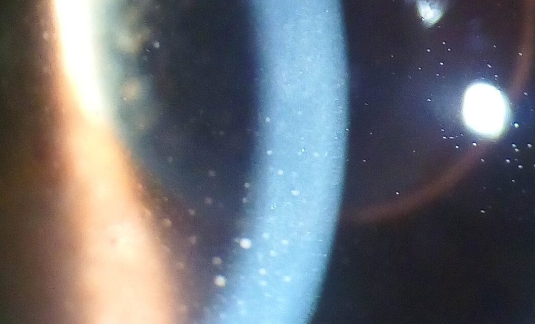
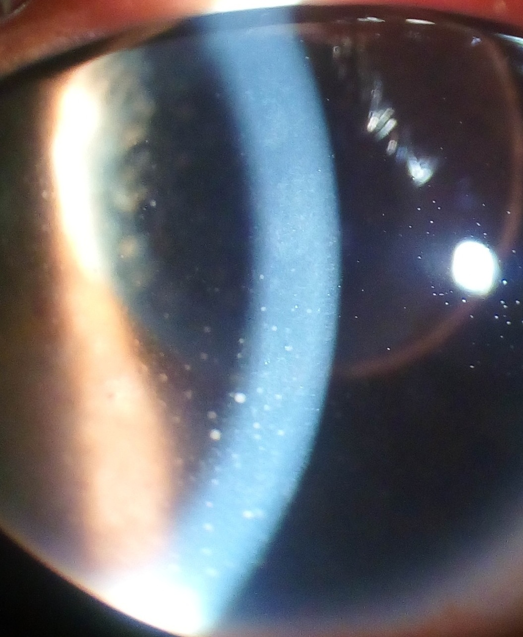
Keratic precipitates are clusters of inflammatory cells that settle onto the corneal endothelium in uveitis, and because different underlying causes of intraocular inflammation tend to produce cells of different types and different characteristic settling patterns, a careful look at their size, shape, color, and distribution at the slit lamp can meaningfully narrow the differential diagnosis before a single laboratory test is ordered.
Why the Pattern Matters
Keratic precipitates form when inflammatory cells circulating in the aqueous humor adhere to the corneal endothelium, and the composition of these cells, whether dominated by large macrophages and epithelioid cells or by smaller lymphocytes and other inflammatory cells, produces visibly different precipitate appearances that correlate, though imperfectly, with the broad category of uveitis present.
Granulomatous Versus Nongranulomatous Patterns
Mutton-Fat Keratic Precipitates
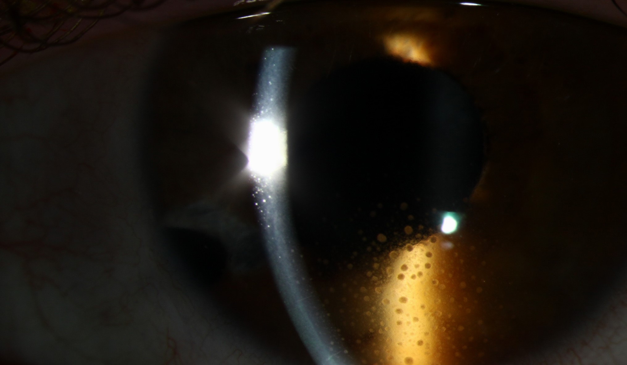
Large, greasy-appearing, yellowish-white precipitates, composed predominantly of macrophages and epithelioid cells, are the hallmark of granulomatous uveitis, and their presence should prompt consideration of the specific causes classically associated with this pattern, including sarcoidosis, tuberculosis, syphilis, and sympathetic ophthalmia, among others.
Fine, Stellate Keratic Precipitates
Small, fine, often stellate (star-shaped) precipitates, in contrast, are typical of nongranulomatous uveitis, seen in conditions such as HLA-B27-associated anterior uveitis and many cases of idiopathic anterior uveitis, and this pattern is also classically associated with Fuchs heterochromic iridocyclitis, a specific, generally mild, chronic uveitis with its own distinct clinical picture.
All-fit Slit-Lamp Adapter
Record and share exactly what you see at the slit lamp. One adapter fits any slit lamp or surgical microscope — and any smartphone.
From Choroida — the team behind this siteDistribution Pattern
- Keratic precipitates classically settle preferentially in the inferior cornea, following the natural convection currents of the aqueous humor, producing a triangular distribution with the base inferiorly, known as Arlt’s triangle
- A diffuse distribution across the entire corneal endothelium, rather than the classic inferior triangular pattern, can be seen in some conditions and is a useful additional descriptive feature
- The specific distribution, combined with the size and color of the precipitates, contributes to the overall clinical impression alongside other examination findings
Additional Descriptive Features
- Fresh versus old precipitates: newly formed precipitates tend to appear more discrete and white, while older precipitates often become more pigmented, irregular, and “crenated” or scalloped in outline as they age, which can provide a rough sense of disease chronicity
- Fine dust-like precipitates: very fine, diffusely scattered precipitates can be seen in some viral anterior uveitis presentations, including those associated with cytomegalovirus, herpes simplex, and varicella-zoster virus
- Coin-shaped or pigmented endothelial lesions: distinct from typical keratic precipitates, though relevant additional findings sometimes present in specific viral anterior uveitis presentations (see viral anterior uveitis)
Using the Pattern in Clinical Practice
Keratic precipitate pattern is one piece of a broader clinical assessment, considered alongside the degree and pattern of anterior chamber inflammation, the presence or absence of posterior synechiae, iris nodules, and any posterior segment findings, and the patient’s demographic and systemic history, rather than being diagnostic on its own.
A granulomatous pattern, for example, appropriately raises the index of suspicion for sarcoidosis or tuberculosis and can help focus an otherwise broad systemic laboratory workup, while a fine, stellate, nongranulomatous pattern in a young patient with recurrent unilateral episodes might point more toward an HLA-B27-associated process.
Documentation
Careful, specific documentation of keratic precipitate size, color, distribution, and any change over serial visits is valuable both for narrowing the differential diagnosis at presentation and for objectively tracking response to anti-inflammatory treatment over the course of follow-up.
Clinical Significance
Recognizing and accurately describing keratic precipitate patterns is a low-cost, immediately available piece of diagnostic information gathered at the very first slit-lamp examination of a patient with uveitis, and while it never replaces a complete history, examination, and targeted laboratory workup, it meaningfully shapes how that broader workup is prioritized and interpreted.


Document what you see
Two smartphone imaging tools built for everyday clinic use — one for the slit lamp, one for the fundus.
From Choroida — the team behind this siteReferences
- Herbort CP, Rao NA, Mochizuki M. International criteria for the diagnosis of ocular sarcoidosis. Ocul Immunol Inflamm. 2009;17:160-169.
- Jabs DA, Nussenblatt RB, Rosenbaum JT. Standardization of uveitis nomenclature for reporting clinical data. Am J Ophthalmol. 2005;140:509-516.
- Bloch-Michel E, Nussenblatt RB. International Uveitis Study Group recommendations for the evaluation of intraocular inflammatory disease. Am J Ophthalmol. 1987;103:234-235.
- Rodriguez A, Calonge M, Pedroza-Seres M, et al. Referral patterns of uveitis in a tertiary eye care center. Arch Ophthalmol. 1996;114:593-599.
