
Conjunctival concretions are small, hard, chalky-white or yellowish deposits that form within the substance of the palpebral conjunctiva, and finding them requires the one extra step that separates a complete external eye examination from an incomplete one: actually everting the eyelid, since these deposits sit precisely where a standard, non-everted examination will never reveal them.
What Concretions Are
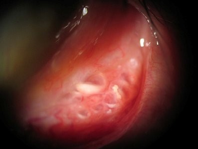
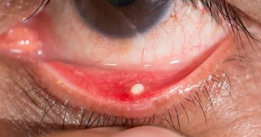
Concretions represent small collections of calcified epithelial debris and cellular material that become trapped within the crypts and folds of the palpebral conjunctiva, most often the inferior tarsal conjunctiva, forming discrete, firm, chalky deposits embedded within the conjunctival tissue itself.
Why Eyelid Eversion Matters
Because concretions form specifically within the tarsal (eyelid-lining) conjunctiva, they are entirely invisible on a standard examination that looks only at the bulbar conjunctiva and the exposed palpebral conjunctiva visible without eyelid manipulation, which is exactly why any patient with a persistent, otherwise unexplained foreign body sensation deserves a deliberate lower and, when appropriate, upper eyelid eversion as part of a complete workup, rather than assuming the sensation must originate from the visible ocular surface alone.
All-fit Slit-Lamp Adapter
Record and share exactly what you see at the slit lamp. One adapter fits any slit lamp or surgical microscope — and any smartphone.
From Choroida — the team behind this siteAssociations and Risk Factors
- Chronic conjunctivitis or blepharoconjunctivitis of any cause, which promotes the epithelial debris accumulation that can eventually calcify into visible concretions
- Advancing age, with a general increase in prevalence
- Chronic dry eye and meibomian gland dysfunction
- Prior ocular surface inflammation or trauma
- Concretions can also occur in otherwise entirely healthy conjunctiva without any identifiable predisposing condition
Clinical Presentation
- Most concretions are entirely asymptomatic, discovered incidentally during eyelid eversion performed for another reason
- When symptomatic, they typically cause a foreign body sensation, mild irritation, or a sense of grittiness, particularly when a concretion has begun to erode through the overlying conjunctival epithelium and creates surface irregularity that can abrade the cornea with blinking
- Multiple concretions are common, sometimes numerous small deposits scattered across the tarsal conjunctiva
- On examination, they appear as discrete, small, firm, white to yellow subconjunctival deposits, generally easily distinguished from other tarsal conjunctival findings by their characteristic chalky appearance and firm consistency on gentle palpation with a cotton-tip applicator
When Concretions Cause Symptoms
The key mechanism by which an otherwise inert, asymptomatic concretion becomes symptomatic is erosion through the overlying conjunctival epithelium, exposing the hard, calcified deposit directly to the ocular surface, where it can then mechanically abrade the cornea with each blink, producing punctate epithelial staining on the cornea corresponding to the position of the exposed concretion.
Management
Asymptomatic Concretions
Most concretions require no treatment at all, since they remain covered by intact conjunctival epithelium and cause no symptoms, and simple observation is entirely appropriate for incidentally discovered, asymptomatic deposits.
Symptomatic Concretions
- For concretions causing genuine foreign body sensation or corneal irritation, removal can be performed at the slit lamp under topical anesthesia, typically using a fine needle or similar instrument to gently elevate and extract the deposit from the conjunctival tissue
- The procedure is generally quick and well tolerated, and multiple concretions can often be addressed in a single sitting if several are contributing to symptoms
- Lubrication is used afterward to support comfort and healing of the small resulting conjunctival defect
Addressing Underlying Contributing Factors
When concretions are associated with chronic conjunctivitis, dry eye, or meibomian gland dysfunction, addressing these underlying conditions can help reduce the likelihood of new concretion formation over time, though this is a secondary consideration rather than the primary treatment for existing, already-formed deposits.
Prognosis
Conjunctival concretions, whether observed or removed, have an excellent prognosis, with removal of symptomatic deposits typically providing prompt and complete symptomatic relief.
Recognizing this common, often overlooked finding, simply by remembering to evert the eyelid during evaluation of unexplained ocular surface irritation, prevents both missed diagnoses and the unnecessary, unproductive workups that can follow when a straightforward, easily treatable cause of foreign body sensation goes unrecognized.


Document what you see
Two smartphone imaging tools built for everyday clinic use — one for the slit lamp, one for the fundus.
From Choroida — the team behind this siteReferences
- Duke-Elder S. System of Ophthalmology. Vol 8. St. Louis: CV Mosby; 1965.
- Sandinha MT, Zaher SS, Roberts F, Devlin H, Dhillon B. Superior tarsal conjunctival concretions: an insight into the aetiology and pathogenesis. Br J Ophthalmol. 2009;93:1329-1330.
- Bowling B. Kanski’s Clinical Ophthalmology: A Systematic Approach. 8th ed. Edinburgh: Elsevier; 2016.
- Sherman SW, Kern H. Conjunctival calcification. Trans Ophthalmol Soc U K. 1978;98:157-159.
