Not every red, painful eye is a corneal or conjunctival problem — sometimes the inflammation is in the wall of the eye itself.
Scleritis is inflammation of the sclera, and it is one of the few ocular surface conditions that regularly signals serious systemic disease.
The pain it produces is different from almost anything else in ophthalmology — deep, boring, and often severe enough to wake a patient from sleep.
Misdiagnosing it as simple conjunctivitis or episcleritis delays both symptom relief and, more importantly, systemic workup.
Roughly half of scleritis cases are linked to an underlying autoimmune or vasculitic disease, and necrotizing forms carry an even higher systemic association.
Recognizing scleritis promptly — and correctly separating it from its much more benign look-alike, episcleritis — genuinely changes outcomes.
What Is Scleritis?
Scleritis is inflammation of the sclera, the tough, collagenous outer coat of the eye, that can threaten both ocular integrity and vision.
It is classified by location and severity:
- Anterior diffuse scleritis — widespread inflammation, the most common and generally the mildest form
- Anterior nodular scleritis — a discrete, immobile, tender inflammatory nodule
- Necrotizing scleritis with inflammation — the most severe and destructive form, with active thinning of the sclera
- Necrotizing scleritis without inflammation (scleromalacia perforans) — painless scleral thinning, classically seen in longstanding rheumatoid arthritis
- Posterior scleritis — inflammation of the sclera behind the equator, which can be easy to miss without imaging
Necrotizing disease carries by far the highest risk of an underlying systemic condition and the greatest threat to the eye itself.
Epidemiology
Scleritis is far less common than episcleritis but carries substantially more clinical weight.
- It typically presents in the fourth to sixth decades of life
- Women are affected more often than men in most reported series
- Roughly half of all scleritis cases are associated with an underlying systemic autoimmune or vasculitic disease
- Necrotizing scleritis carries the highest systemic association of any subtype, reported in the range of 70-95% of cases
Because the systemic association is so high, every new diagnosis of scleritis should prompt at least a basic review for underlying autoimmune disease.
All-fit Slit-Lamp Adapter
Record and share exactly what you see at the slit lamp. One adapter fits any slit lamp or surgical microscope — and any smartphone.
From Choroida — the team behind this sitePathophysiology
Scleritis is fundamentally an immune-mediated process rather than an infectious one in the great majority of cases.
- Immune complex deposition and vasculitis within scleral and episcleral vessels drive the inflammatory process
- Inflammatory cell infiltration and cytokine release damage scleral collagen and the resident fibroblasts
- In necrotizing disease, occlusive vasculitis compromises blood flow to the sclera itself
- Progressive collagen destruction leads to scleral thinning, which can progress to visible uveal show or, rarely, perforation
Because the sclera is relatively avascular and slow to heal, established scleral thinning from necrotizing disease is often permanent even once inflammation is controlled.
Risk Factors
Systemic Associations
- Rheumatoid arthritis — the most common systemic disease associated with scleritis
- Granulomatosis with polyangiitis (GPA) — the most common vasculitis associated with scleritis, and a classic cause of necrotizing disease
- Other systemic vasculitides, including relapsing polychondritis and polyarteritis nodosa
- Inflammatory bowel disease and systemic lupus erythematosus, less commonly
Other Risk Factors
- Prior ocular surgery, which can trigger surgically induced necrotizing scleritis
- Infectious causes (bacterial, viral, or fungal), which are uncommon but must be actively excluded before immunosuppression
- Middle age, given the peak incidence in the fourth to sixth decades
Identifying a systemic trigger changes management directly — infectious causes need targeted anti-microbial therapy instead of immunosuppression.
Clinical Presentation
Symptoms
- Deep, boring, severe eye pain, often radiating to the brow, temple, or jaw
- Pain frequently worse at night and severe enough to wake the patient from sleep
- Marked tenderness to gentle palpation of the globe through closed lids
- Photophobia and tearing, particularly when the cornea is also involved
- Gradual, sometimes painless vision loss in posterior scleritis
Examination Findings
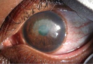
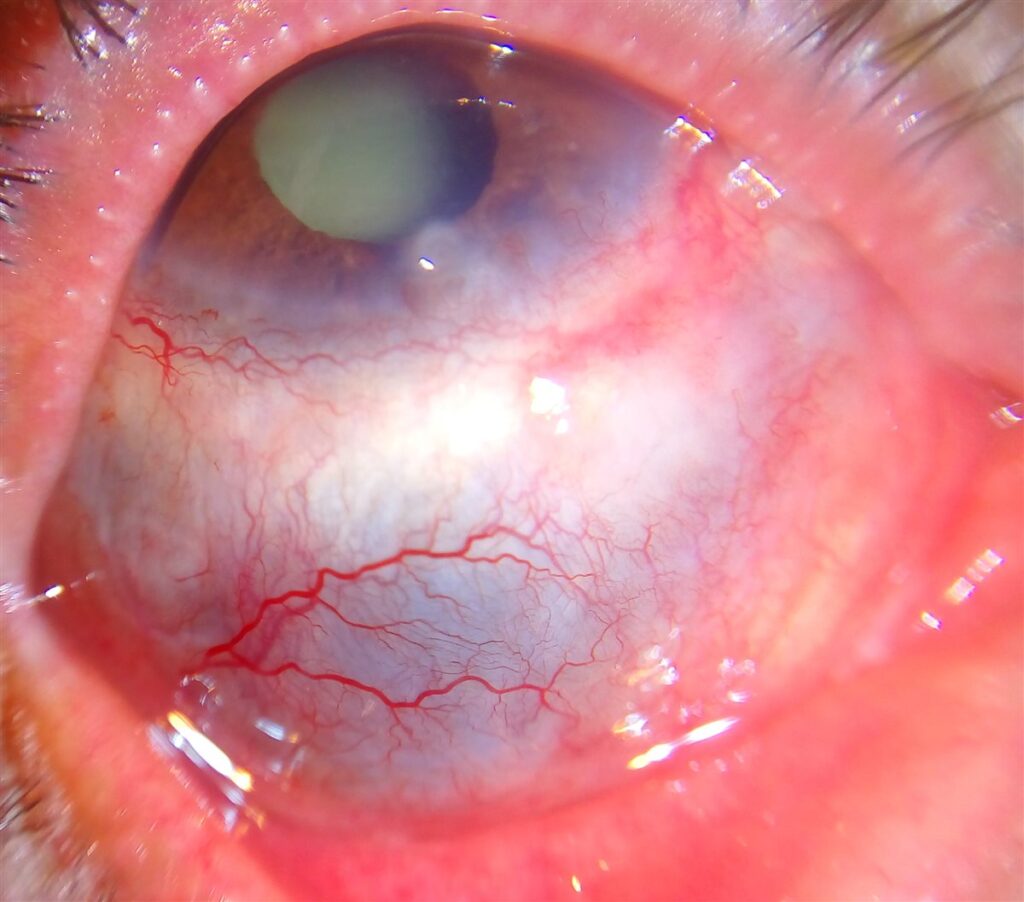
- Deep, violaceous or bluish-red engorgement of the scleral and deep episcleral vascular plexus
- Vessels that do not blanch with topical phenylephrine, unlike episcleritis
- Scleral edema, nodules, or, in longstanding necrotizing disease, visible bluish-gray thinning that lets the underlying uveal pigment show through
- Associated peripheral ulcerative keratitis in some necrotizing cases
- Anterior chamber inflammation or, in posterior scleritis, choroidal folds and disc edema on fundus exam
The phenylephrine blanching test is the single most useful bedside maneuver: episcleral vessels blanch, scleritis vessels do not.
Diagnostic Evaluation
Clinical Examination
- Slit-lamp exam to characterize the pattern of vascular engorgement and check for scleral thinning or nodules
- Topical phenylephrine or neosynephrine blanching test to distinguish scleritis from episcleritis
- Careful palpation for tenderness, which is characteristic of scleritis and unusual in episcleritis
Imaging
- B-scan ultrasonography to detect posterior scleritis, classically showing scleral thickening and the “T-sign” from fluid in Tenon’s space
- Anterior segment OCT can help characterize scleral and episcleral involvement in select cases
Systemic Workup
- Complete blood count, inflammatory markers (ESR, CRP), and rheumatoid factor
- ANCA testing when vasculitis, particularly GPA, is suspected
- Targeted infectious workup (e.g., syphilis serology, tuberculosis screening) before starting systemic immunosuppression
- Referral to rheumatology when a systemic association is identified or strongly suspected
Scleritis should always be treated as a potential sentinel sign of systemic disease until proven otherwise, not managed as an isolated eye problem.
Differential Diagnosis
Conditions that can be confused with scleritis include:
- Episcleritis — milder, vessels blanch with phenylephrine, minimal to no tenderness, generally self-limited
- Conjunctivitis — diffuse conjunctival injection with discharge, but without the deep violaceous hue or globe tenderness
- Infectious scleritis — often follows surgery or trauma, may show a discrete abscess or infiltrate, requires microbiological workup
- Anterior uveitis — pain and photophobia can overlap, but redness is typically circumlimbal rather than the deep scleral pattern of scleritis
- Orbital cellulitis or inflammation — associated proptosis or restricted motility not typical of isolated scleritis
Distinguishing scleritis from episcleritis at the bedside is the single highest-yield step, since it determines whether systemic workup is warranted at all.
Management
Mild to Moderate Non-Necrotizing Disease
- Oral non-steroidal anti-inflammatory drugs (NSAIDs) are frequently effective first-line therapy
- Topical corticosteroids can provide adjunctive symptomatic relief but do not treat the disease adequately on their own
Moderate to Severe or Necrotizing Disease
- Systemic corticosteroids form the backbone of treatment for disease not controlled with NSAIDs alone
- Steroid-sparing immunosuppressive agents (e.g., methotrexate, mycophenolate, cyclophosphamide) are used for necrotizing disease, recurrent disease, or when a systemic vasculitis is identified
- Biologic therapy (e.g., rituximab) is increasingly used for refractory necrotizing scleritis, particularly when associated with GPA
Coordinated Systemic Care
- Close co-management with rheumatology once a systemic association is confirmed or strongly suspected
- Infectious causes must be excluded or specifically treated before committing to long-term immunosuppression
Necrotizing scleritis, in particular, is a marker of systemic disease activity — treating the eye alone without addressing the underlying vasculitis risks both the eye and the patient’s broader health.
Prognosis
Outcomes vary substantially by subtype and by how quickly systemic disease, when present, is brought under control.
- Diffuse and nodular anterior scleritis generally carry a favorable visual prognosis with appropriate treatment
- Necrotizing scleritis carries the highest risk of scleral thinning, ocular complications, and vision loss
- Poor control of an underlying systemic vasculitis is associated with both worse ocular outcomes and increased systemic morbidity
Long-term outcome in scleritis is inseparable from control of the systemic disease driving it, particularly in necrotizing cases.
Would you like to document anterior segment findings with your smartphone?


Document what you see
Two smartphone imaging tools built for everyday clinic use — one for the slit lamp, one for the fundus.
From Choroida — the team behind this siteReferences
- Wieringa WG, Wieringa JE, ten Dam-van Loon NH, et al. Current Approach for the Diagnosis and Management of Noninfective Scleritis. Ocular Immunology and Inflammation. 2023.
- Sainz de la Maza M, Molina N, Gonzalez-Gonzalez LA, et al. Clinical characteristics of a large cohort of patients with scleritis and episcleritis. Ophthalmology. 2012.
- Scleritis. StatPearls, NCBI Bookshelf.
- Watson PG, Hayreh SS. Scleritis and episcleritis. British Journal of Ophthalmology. 1976.
- Scleritis. EyeWiki, American Academy of Ophthalmology.
Test yourself
A few questions straight from this article.
-
Scleritis is inflammation of which ocular structure?
Scleritis is inflammation of the sclera, the tough collagenous outer coat, and it can threaten both the structural integrity of the eye and vision. -
Which pain pattern is characteristic of scleritis?
The pain is deep and boring, often radiates to the brow, temple or jaw, is typically worse at night, and is frequently severe enough to wake the patient from sleep. -
Roughly what proportion of scleritis cases are linked to systemic autoimmune or vasculitic disease?
Roughly half of all scleritis cases carry an underlying systemic autoimmune or vasculitic association, which is why every new diagnosis warrants at least a basic systemic review. -
Necrotizing scleritis carries a systemic disease association reported in which range?
Necrotizing scleritis has the highest systemic association of any subtype, reported in the range of 70-95% of cases, along with the greatest threat to the eye. -
Which bedside test best separates scleritis from episcleritis?
The phenylephrine blanching test is the single most useful bedside manoeuvre: episcleral vessels blanch with the drop, whereas the deep vessels of scleritis do not. -
Which systemic disease is most commonly associated with scleritis?
Rheumatoid arthritis is the most common systemic disease associated with scleritis, and longstanding disease is the classic setting for painless scleromalacia perforans. -
Which vasculitis is most commonly associated with scleritis and classically causes necrotizing disease?
Granulomatosis with polyangiitis is the vasculitis most often linked to scleritis and a classic cause of necrotizing disease, which is why ANCA testing is warranted when vasculitis is suspected. -
Which investigation detects posterior scleritis by demonstrating the T-sign?
B-scan ultrasonography classically shows scleral thickening and the T-sign from fluid in Tenon's space, which is how posterior scleritis is confirmed. -
What is frequently effective first-line therapy for mild to moderate non-necrotizing scleritis?
Oral NSAIDs are frequently effective first-line; topical corticosteroids give adjunctive symptomatic relief but do not adequately treat the disease on their own. -
Which step must precede long-term systemic immunosuppression for scleritis?
Infectious scleritis is uncommon but must be actively excluded or specifically treated before committing a patient to long-term immunosuppression.

Writes and reviews the clinical articles on Educate. Every post is checked against peer-reviewed sources before it goes live.