
Scleritis classification into anatomical location and inflammatory pattern is not just descriptive labeling; the specific type of scleritis a patient has strongly predicts both the likelihood of finding an associated systemic autoimmune disease and the risk of vision-threatening complications, which is why the classification shapes the workup from the very first visit.
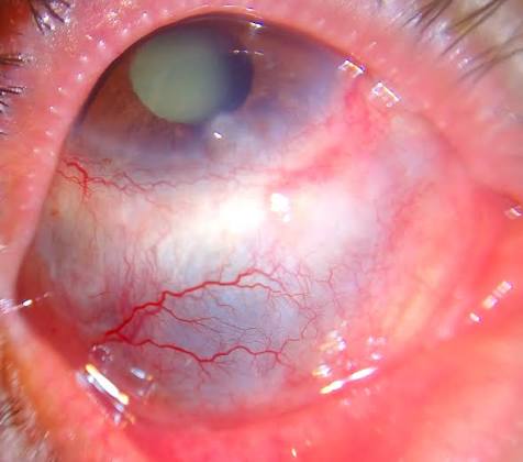
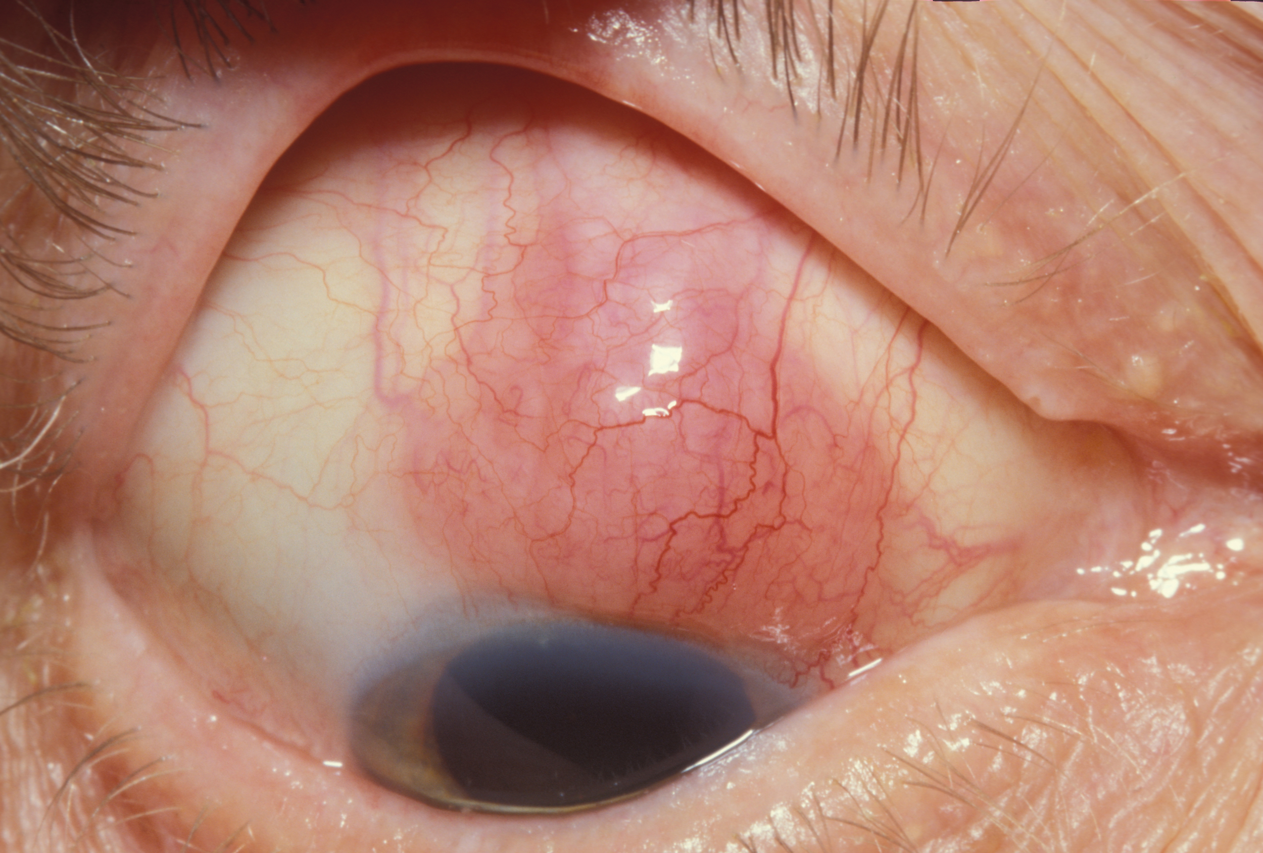
Anterior Scleritis
Anterior scleritis is the most common form and is subdivided by clinical appearance.
- Diffuse anterior scleritis: widespread scleral inflammation and redness, the most common and generally the most benign pattern
- Nodular anterior scleritis: one or more discrete, immobile, tender nodules of scleral inflammation
- Necrotizing anterior scleritis with inflammation: the most severe and destructive pattern, with thinning and potential perforation of the sclera, strongly associated with systemic autoimmune disease
- Necrotizing anterior scleritis without inflammation (scleromalacia perforans): a distinct, painless variant seen almost exclusively in long-standing rheumatoid arthritis, where scleral thinning progresses with little apparent inflammation, sometimes to the point of visible uveal show through the thinned sclera (see scleromalacia perforans)
Posterior Scleritis
Posterior scleritis involves the sclera behind the equator and is more likely to be missed clinically, since the eye can look relatively white and quiet externally while significant inflammation and pain are present.
- Pain, sometimes severe, out of proportion to external findings
- Proptosis, restricted motility, and choroidal folds or exudative retinal detachment from posterior involvement
- B-scan ultrasonography showing scleral and episcleral thickening, sometimes with a characteristic “T-sign” from fluid in the sub-Tenon space
Posterior scleritis is frequently underdiagnosed because of the deceptively quiet external appearance, and it should be considered in any patient with unexplained ocular pain, especially with proptosis or restricted movement (see posterior scleritis).
All-fit Slit-Lamp Adapter
Record and share exactly what you see at the slit lamp. One adapter fits any slit lamp or surgical microscope — and any smartphone.
From Choroida — the team behind this siteWhy the Pattern Matters for the Workup
Necrotizing scleritis, whether anterior with inflammation or the painless scleromalacia perforans variant, carries the strongest association with systemic autoimmune disease and the highest risk of ocular and even life-threatening systemic complications, and it warrants the most thorough and urgent systemic evaluation.
Diffuse and nodular anterior scleritis are more often idiopathic but still carry a meaningful minority risk of systemic association and deserve at least a baseline autoimmune and infectious screen.
Systemic Associations
- Rheumatoid arthritis, one of the most common associations, particularly with necrotizing disease and scleromalacia perforans
- Granulomatosis with polyangiitis, strongly linked to necrotizing scleritis and requiring urgent recognition given its potential severity (see granulomatosis with polyangiitis)
- Relapsing polychondritis
- Systemic lupus erythematosus and other connective tissue diseases
- Infectious causes, including herpes zoster, tuberculosis, and syphilis, particularly relevant after ocular surgery or trauma
- Gout, though less common as a cause of true scleritis
Workup
- Complete blood count, inflammatory markers (ESR, CRP)
- Rheumatoid factor and anti-CCP antibodies
- ANCA testing, particularly for suspected granulomatosis with polyangiitis
- ANA and other lupus-related serology when indicated
- Syphilis and tuberculosis testing, especially with atypical or necrotizing presentations
- Chest imaging when systemic vasculitis or sarcoidosis is suspected
The extent of workup is tailored to the clinical pattern: a mild first episode of diffuse anterior scleritis in an otherwise healthy patient may warrant a more limited initial screen, while necrotizing disease warrants prompt, thorough systemic evaluation.
Management Implications by Type
Diffuse and nodular scleritis often respond to oral NSAIDs or systemic corticosteroids, while necrotizing scleritis typically requires systemic corticosteroids together with steroid-sparing immunosuppression, coordinated with rheumatology given the strong systemic disease association (see necrotizing scleritis).
Prognosis
Diffuse and nodular anterior scleritis generally carry a favorable ocular prognosis with appropriate treatment.
Necrotizing scleritis carries a real risk of scleral perforation, vision loss, and, when associated with systemic vasculitis, increased mortality if the underlying systemic disease is not adequately treated, which is the central reason classification is not just an academic exercise.


Document what you see
Two smartphone imaging tools built for everyday clinic use — one for the slit lamp, one for the fundus.
From Choroida — the team behind this siteReferences
- Watson PG, Hayreh SS. Scleritis and episcleritis. Br J Ophthalmol. 1976;60:163-191.
- Sainz de la Maza M, Foster CS, Jabbur NS. Scleritis associated with systemic vasculitic diseases. Ophthalmology. 1995;102:687-692.
- Wieringa WG, Wieringa JE, ten Dam-van Loon NH, Los LI. Visual outcome, treatment results, and prognostic factors in patients with scleritis. Ophthalmology. 2013;120:379-386.
- Akpek EK, Thorne JE, Qazi FA, Do DV, Jabs DA. Evaluation of patients with scleritis for systemic disease. Ophthalmology. 2004;111:501-506.
