
Iris mammillations produce a distinctive, uniformly nodular, velvety texture across the iris surface, and recognizing this specific pattern matters mainly because it differs so clearly from the focal, irregular growth pattern of a worrisome iris lesion, letting a clinician confidently reassure a patient about a finding that might otherwise look alarming at first glance.
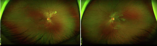
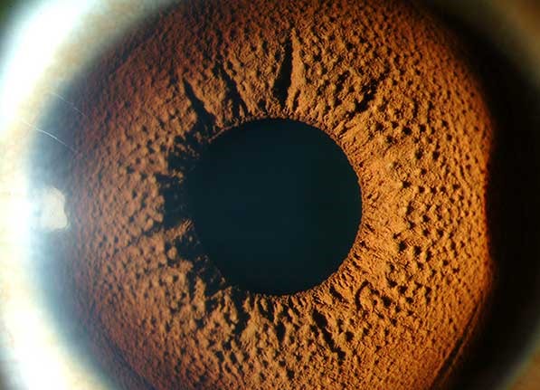
What Mammillations Look Like
Rather than a single, focal pigmented lesion, iris mammillations present as small, regularly spaced, uniform nodular or villiform projections covering some or all of the iris surface, giving the iris a velvety or cobblestone appearance that is symmetric and diffuse rather than localized.
This diffuse, symmetric surface change is fundamentally different in character from a nevus or melanoma, which present as a discrete, localized area of altered pigmentation or elevation against an otherwise normal-appearing surrounding iris.
Associations
- Isolated, congenital iris mammillations occur without any other associated finding in some individuals, representing a benign anatomic variant of normal iris surface architecture
- Oculodermal melanocytosis (nevus of Ota), a condition involving increased dermal melanocyte density affecting the periocular skin, sclera, and sometimes the iris and other ocular structures, is a recognized association, and iris mammillations can be a subtle clue to this broader diagnosis when present
- When associated with oculodermal melanocytosis, the affected eye carries an increased lifetime risk of uveal melanoma, which is the primary reason this broader diagnosis, once suspected, warrants ongoing surveillance
All-fit Slit-Lamp Adapter
Record and share exactly what you see at the slit lamp. One adapter fits any slit lamp or surgical microscope — and any smartphone.
From Choroida — the team behind this siteClinical Significance
The main clinical value of recognizing iris mammillations lies in correctly distinguishing this benign, diffuse surface texture change from a genuinely concerning focal iris lesion, avoiding unnecessary anxiety, imaging, or biopsy for a finding that, in isolation, carries no malignant potential of its own.
At the same time, recognizing mammillations as a potential clue to underlying oculodermal melanocytosis, when other supporting features are present, such as increased scleral pigmentation or ipsilateral periocular skin hyperpigmentation, prompts the appropriately more vigilant long-term surveillance that this broader condition warrants, given its association with elevated uveal melanoma risk.
Examination
- Careful slit-lamp examination of the iris surface, assessing whether an observed pigmentary or textural change is diffuse and symmetric (favoring mammillations as a variant) or focal and irregular (favoring a nevus or other discrete lesion requiring its own separate risk assessment)
- Examination for other signs of oculodermal melanocytosis, including scleral pigmentation (often patchy, slate-gray in color) and ipsilateral periocular or facial dermal pigmentation
- Documentation with slit-lamp photography, useful both for the patient’s own record and for any future comparison
Management
Isolated Mammillations
No specific treatment or intervention is needed for isolated iris mammillations without other associated findings, and routine ongoing eye care is appropriate, since this represents a benign anatomic variant rather than a disease process.
When Associated With Oculodermal Melanocytosis
When mammillations occur as part of oculodermal melanocytosis, longer-term surveillance is appropriate given the recognized association with increased uveal melanoma risk in the affected eye, including periodic dilated fundus examination to monitor for the development of a choroidal or other uveal melanocytic lesion over time, coordinated with the patient’s general eye care.
Patient Education
Patients found to have oculodermal melanocytosis benefit from clear education about the modestly increased melanoma risk and the rationale for ongoing surveillance, framed in a way that supports adherence to regular follow-up without causing undue alarm, given that the great majority of affected individuals never develop melanoma.
Prognosis
Isolated iris mammillations, without other associated findings, carry no independent implications for ocular health and require no specific monitoring beyond routine eye care.
When associated with oculodermal melanocytosis, the long-term outlook depends on ongoing surveillance catching any eventual melanocytic transformation early, which is the entire rationale behind recommending sustained, periodic follow-up for these patients rather than a one-time evaluation.


Document what you see
Two smartphone imaging tools built for everyday clinic use — one for the slit lamp, one for the fundus.
From Choroida — the team behind this siteReferences
- Rennie IG. Don’t it make my brown eyes blue–iris colour and related pathology. Eye (Lond). 2012;26:29-42.
- Shields CL, Kaliki S, Livesey M, et al. Association of ocular and oculodermal melanocytosis with the rate of uveal melanoma metastasis: analysis of 7872 consecutive eyes. JAMA Ophthalmol. 2013;131:993-1003.
- Teekhasaenee C, Ritch R, Rutnin U, Leelawongs N. Ocular findings in oculodermal melanocytosis. Arch Ophthalmol. 1990;108:1114-1120.
- Singh AD, De Potter P, Fijal BA, Shields CL, Shields JA, Elston RC. Lifetime prevalence of uveal melanoma in white patients with oculo(dermal) melanocytosis. Ophthalmology. 1998;105:195-198.
Test yourself
A few questions straight from this article.
-
Which iris surface appearance characterizes iris mammillations?
Iris mammillations are small, regularly spaced, uniform nodular or villiform projections covering some or all of the iris, producing a velvety or cobblestone look that is symmetric and diffuse. -
Which pattern on slit-lamp examination favours iris mammillations as a normal variant?
A diffuse, symmetric textural change favours mammillations as a variant, whereas a focal, irregular change favours a nevus or other discrete lesion needing its own risk assessment. -
What is the main clinical value of recognizing iris mammillations?
Correctly identifying this benign diffuse surface texture, rather than mistaking it for a concerning focal lesion, spares the patient unnecessary anxiety, imaging or biopsy for a finding with no malignant potential of its own. -
How are isolated iris mammillations with no other findings best managed?
Isolated iris mammillations represent a benign anatomic variant rather than a disease process, so no specific treatment is needed and routine ongoing eye care is appropriate. -
Which condition is a recognized association of iris mammillations?
Oculodermal melanocytosis, also called nevus of Ota, is a recognized association, and iris mammillations can be a subtle clue to that broader diagnosis. -
What underlies oculodermal melanocytosis?
Oculodermal melanocytosis involves increased dermal melanocyte density affecting the periocular skin, sclera and sometimes the iris and other ocular structures. -
Which risk does oculodermal melanocytosis confer on the affected eye?
When mammillations occur as part of oculodermal melanocytosis, the affected eye carries an increased lifetime risk of uveal melanoma, which is why the broader diagnosis warrants surveillance. -
Which additional signs support a diagnosis of oculodermal melanocytosis?
Examination should look for scleral pigmentation, often patchy and slate-gray, together with ipsilateral periocular or facial dermal pigmentation. -
Which surveillance is recommended when mammillations occur with oculodermal melanocytosis?
Longer-term surveillance including periodic dilated fundus examination monitors for development of a choroidal or other uveal melanocytic lesion over time. -
Which point should counselling about oculodermal melanocytosis convey?
Education should explain the modestly increased melanoma risk and the rationale for follow-up without undue alarm, since the great majority of affected individuals never develop melanoma.
