Anterior segment OCT (AS-OCT, or anterior segment optical coherence tomography) is a non-contact scan that produces cross-sectional images of the cornea, anterior chamber, angle, and iris. It takes seconds, needs no dye or gel, and never touches the eye. Clinicians use it mainly to measure corneal thickness, judge angle width, and see through hazy corneas.
The technique extends optical coherence tomography, most familiar as a posterior-segment tool, to the front of the eye. It fills a different clinical niche than daily retinal scans and competes with ultrasound biomicroscopy for some of the same questions. Newer swept-source platforms image deeper and faster than earlier time-domain systems, so one scan now captures more of the anterior segment.
What the AS OCT Eye Test Involves
The anterior segment is the front third of the eye, running from the cornea through the anterior chamber and iris to the front of the lens. For the AS OCT eye test, the patient rests the chin on a support and looks at a fixation target while the device scans. Each image takes a few seconds.
The scan uses near-infrared light, involves no radiation, and carries no known risk to the eye. Most patients need no drops. Because nothing touches the eye, an anterior segment OCT test suits children, anxious patients, and eyes with fragile surfaces or recent surgery.
You may see the same test written as AS OCT, OCT AS, anterior OCT, or anterior seg OCT, and the printout may be called an AS OCT scan. Some devices are dedicated anterior units, while others are retinal OCT machines fitted with an anterior segment lens or module.
How Anterior Segment OCT Differs From Posterior OCT and UBM
Anterior segment OCT uses the same interferometric principle as posterior OCT, but its imaging parameters suit the shallower depth and different reflective properties of anterior structures rather than the retina. That tuning gives high-resolution images of the cornea and angle in a single, quick session.
Unlike ultrasound biomicroscopy (UBM), which needs a water bath or gel coupling and direct probe contact, anterior segment OCT is entirely non-contact. That makes it faster and more comfortable, particularly for anxious or pediatric patients, and it avoids any risk of abrading the surface.
UBM still outperforms anterior segment OCT for structures behind the iris, such as the ciliary body, because light cannot penetrate the iris pigment epithelium the way sound waves can. The two tools are complementary rather than interchangeable, and the structure that needs to be seen should drive the choice.
Fundus Explorer Pro
Photograph the retinal findings described here with the phone already in your pocket — 22 D optics and built-in illumination in one handheld unit.
From Choroida — the team behind this siteClinical Uses of Anterior Segment OCT
- Angle assessment: a cross-sectional, quantifiable view of angle width and configuration that complements gonioscopy, especially for screening and for patients who cannot tolerate a goniolens
- Corneal pachymetry mapping across the whole cornea, more detailed than a single central ultrasound reading
- Pre-refractive surgery planning: corneal thickness and shape, to help exclude eyes at higher risk of post-surgical ectasia
- Corneal graft monitoring after keratoplasty, including graft apposition, interface fluid, and early signs of rejection or detachment
- Imaging hazy or edematous corneas where the slit-lamp view is too degraded for direct assessment
- Checking the position of an anterior chamber intraocular lens and its relation to the angle
Angle Assessment With Anterior Chamber OCT
Anterior chamber OCT gives an objective, reproducible cross-sectional image of the angle that can be captured quickly and read without the hands-on skill gonioscopy demands. It is useful as a screening tool for angle closure risk in populations, and for documenting angle configuration over time in a patient with narrowing.
It also pairs well with the Van Herick test, which estimates limbal chamber depth at the slit lamp. When the Van Herick grade suggests a shallow periphery, OCT can measure the chamber and angle directly and store the result for comparison.
Gonioscopy remains necessary for functional assessment. It allows dynamic indentation and direct viewing of pigmentation and peripheral anterior synechiae, which anterior segment OCT alone does not reliably capture. Use OCT for fast, reproducible, quantitative screening and gonioscopy for the functional and pigmentary detail that only direct visualization provides.
Anterior Segment OCT Interpretation
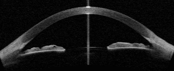
Start by checking image quality and orientation, then locate the scleral spur, which anchors most angle measurements. From there, a reader typically reviews these measures:
- Angle opening distance (AOD) and trabecular-iris space area (TISA), measured from the scleral spur
- Anterior chamber depth and width
- Lens vault, the height of the anterior lens surface above the line joining the two scleral spurs
- Iris thickness, curvature, and any contact with the trabecular meshwork or lens
A narrow angle with iris contact against the trabecular meshwork suggests appositional closure, and a shallow chamber with a high lens vault points toward a lens-related mechanism. Lighting changes pupil size and angle width, so compare scans taken under the same conditions.
Corneal maps show thickness across the whole cornea. Focal thinning or asymmetry raises suspicion for keratoconus, and the pattern should be confirmed with corneal topography or tomography. Always correlate the scan with the slit-lamp exam rather than reading it alone.
Imaging an Opaque Cornea
When a cornea is too hazy or edematous for a clear slit-lamp view, anterior segment OCT can often still produce a usable cross-sectional image, because it relies on a different physical principle than direct inspection. That makes it valuable for assessing corneal thickness, graft-host apposition, or Descemet membrane status before the cornea clears.
Intraoperative OCT, integrated into some operating microscopes, brings the same benefit into surgery. It lets the surgeon check graft orientation, residual Descemet membrane fragments, and dissection depth in real time, as in deep anterior lamellar keratoplasty, instead of relying on visual inspection alone.
Other Anterior Segment Imaging Tools
AS-OCT is one of several anterior segment imaging methods. Scheimpflug tomography maps the front and back corneal surfaces and is the usual companion for ectasia screening. Specular microscopy counts endothelial cells, and UBM images the ciliary body and zonules.
Slit-lamp photography documents surface findings, and in vivo confocal microscopy shows cellular detail. Each answers a different question, so choose the test by the clinical problem and add AS-OCT when a cross-sectional, non-contact view of anterior structures will change management.
Limitations of AS OCT
Image quality depends on corneal clarity, and very dense opacities or significant surface irregularity can degrade the scan enough to limit its usefulness. Structures posterior to the iris are poorly seen because light cannot penetrate the iris pigment epithelium, which keeps UBM clinically relevant.
Interpretation requires familiarity with the artifacts and normal variants of anterior segment scans, which differ enough from posterior segment OCT that experience with one does not automatically transfer to the other. Angle measurements also depend on correct scleral spur placement, which software sometimes gets wrong.
Cost and access remain practical constraints, since dedicated or add-on anterior segment OCT is not available everywhere the way slit-lamp examination and gonioscopy are. A scan also supplements the clinical exam and does not replace it.


Document what you see
Two smartphone imaging tools built for everyday clinic use — one for the slit lamp, one for the fundus.
From Choroida — the team behind this siteReferences
- Radhakrishnan S, Rollins AM, Roth JE, et al. Real-time optical coherence tomography of the anterior segment at 1310 nm. Archives of Ophthalmology.
- American Academy of Ophthalmology. Basic and Clinical Science Course, Section 8: External Disease and Cornea.
- Nolan WP, See JL, Chew PT, et al. Detection of primary angle closure using anterior segment optical coherence tomography in Asian eyes. Ophthalmology.
Test yourself
A few questions straight from this article.
-
What does anterior segment optical coherence tomography image?
Anterior segment OCT applies the interferometric principle of posterior OCT to the front of the eye, producing non-contact cross-sectional images of cornea, angle and anterior chamber. -
Which practical difference separates anterior segment OCT from ultrasound biomicroscopy?
Ultrasound biomicroscopy needs a water bath or gel and direct probe contact with the eye, while anterior segment OCT touches nothing, making it faster and easier for anxious or paediatric patients. -
Which structure does ultrasound biomicroscopy image better than anterior segment OCT?
Light cannot penetrate the iris pigment epithelium the way sound waves can, so structures behind the iris remain the territory of ultrasound biomicroscopy. -
What advantage do swept-source platforms bring to anterior segment OCT over earlier time-domain systems?
Swept-source platforms have considerably improved the depth and speed of anterior segment imaging, widening what a single scan can reliably capture. -
How does anterior segment OCT pachymetry improve on ultrasound pachymetry?
Anterior segment OCT produces a thickness map over the entire cornea, which is far more detailed than the single central reading a standard ultrasound pachymeter gives. -
What is anterior segment OCT used for when planning refractive surgery?
Characterising corneal thickness and shape before refractive surgery helps identify the eyes most likely to develop ectasia after the procedure. -
Why does gonioscopy remain necessary even when anterior segment OCT imaging of the angle is available?
Only direct visualisation allows dynamic indentation and reliable assessment of angle pigmentation and peripheral anterior synechiae, which anterior segment OCT does not capture. -
What can anterior segment OCT assess in an eye whose cornea is too oedematous for a slit-lamp view?
Because it relies on a different physical principle from direct inspection, anterior segment OCT often still yields a usable cross-section when the cornea is hazy, guiding the surgeon until it clears. -
What does intraoperative anterior segment OCT built into an operating microscope allow a surgeon to confirm?
Real-time imaging during surgery lets the surgeon verify structures such as graft orientation or leftover Descemet fragments rather than relying on visual inspection alone. -
Which interpretive limitation applies to anterior segment OCT for a clinician experienced in posterior OCT?
Anterior segment scans carry their own artifacts and normal variants, and familiarity with posterior segment OCT does not automatically transfer to reading them correctly.

Writes and reviews the clinical articles on Educate. Every post is checked against peer-reviewed sources before it goes live.