
Conjunctival autografting is the technique that changed pterygium surgery from a procedure with a notoriously high recurrence rate into one with results good enough to be considered definitive in most patients.
Before its adoption, bare sclera excision alone left recurrence rates high enough that many surgeons and patients viewed pterygium surgery as something that would likely need to be repeated. Autografting did not eliminate recurrence, but it brought it down substantially, and that difference is the entire rationale for the extra surgical step involved.
Why the Graft Matters
A pterygium is a fibrovascular growth of conjunctival and subconjunctival tissue that invades the cornea, typically from the nasal limbus, and surgical excision alone leaves bare sclera exposed at the excision site.
Bare sclera left uncovered is what drives high recurrence after simple excision: exposed, inflamed tissue provides exactly the environment in which fibrovascular tissue regrows and reinvades the cornea.
Covering that bare sclera with a free conjunctival autograft, harvested from another part of the eye, provides a physical and biological barrier that substantially lowers this recurrence risk compared with leaving the area to heal on its own.
Surgical Technique
The pterygium head is dissected off the cornea, and the body of the lesion is excised from the conjunctiva and underlying Tenon capsule down to bare sclera, with care taken to remove the fibrovascular tissue thoroughly to reduce the substrate for recurrence.
A free conjunctival graft, usually harvested from the superior bulbar conjunctiva of the same eye where it is least noticeable, is then sized to match the defect and transferred to cover the bare sclera.
The graft is secured either with fine sutures or with fibrin tissue glue, orientated so that the limbal edge of the graft is placed at the limbus, preserving the normal anatomical relationship and supporting limbal stem cell function at the site.
All-fit Slit-Lamp Adapter
Record and share exactly what you see at the slit lamp. One adapter fits any slit lamp or surgical microscope — and any smartphone.
From Choroida — the team behind this siteSuture Versus Glue Fixation
Suture fixation is well established, widely available, and does not depend on specialised adhesive products, but sutures themselves can cause postoperative discomfort, granuloma formation, and require removal or gradual dissolution.
Fibrin glue fixation generally causes less postoperative discomfort and reduces operating time, with comparable recurrence rates to sutures in most comparative studies, though it is more costly and not universally available.
The choice between the two often comes down to surgeon experience, cost, and local availability of tissue glue rather than a clear superiority of one technique over the other for recurrence prevention specifically.
Alternatives and Adjuncts
Amniotic membrane transplantation can be used instead of conjunctival autograft, particularly when conjunctival tissue is limited, such as in recurrent pterygium or a patient who may need future glaucoma surgery where conjunctival tissue should be preserved.
Adjunctive mitomycin C is used in some recurrent or higher-risk cases to further reduce fibroblast proliferation and recurrence risk, though it carries its own risks, including scleral thinning and delayed healing, and is generally reserved for selected cases rather than used routinely.
Bare sclera excision without any graft or adjunct is now considered inferior for recurrence prevention and is generally reserved for situations where graft harvesting is not feasible.
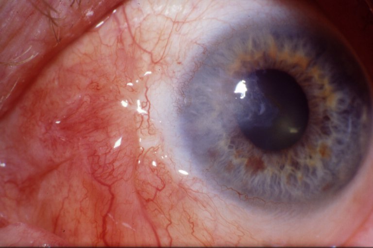
Postoperative Course and Appearance
Conjunctival injection and mild discomfort are expected in the early postoperative period as the graft vascularises and integrates with the surrounding tissue, generally settling over several weeks.
A residual corneal opacity or scar can remain at the site of the original pterygium head, particularly with larger or more advanced lesions, even when the surgery itself has successfully prevented recurrence. This is worth setting expectations for preoperatively, since a technically successful, non-recurrent result does not always mean a cosmetically perfect one, especially in eyes with more extensive corneal involvement before surgery.
Complications
- Graft failure or retraction, occasionally requiring a further procedure to secure or replace the graft
- Granuloma formation at suture sites, when sutures rather than glue are used
- Recurrence, still possible despite grafting, though at a substantially lower rate than with bare sclera excision alone
- Dellen formation adjacent to the graft from localised tear film disruption, usually resolving with lubrication
- Symblepharon, adhesion between the conjunctiva and the globe, uncommon with careful technique but more of a risk in extensive or recurrent surgery
Prognosis
Recurrence rates after conjunctival autografting are substantially lower than after bare sclera excision alone, and this difference is well established across multiple comparative studies, making autografting the accepted standard for primary pterygium surgery in most settings.
Recurrent pterygium, when it does occur, tends to be more aggressive and harder to manage than the original lesion, which is part of why getting the first surgery right, with appropriate graft coverage, matters more than it might first appear.
Visual outcome depends heavily on how much corneal involvement and astigmatism the pterygium had caused before surgery. Lesions caught and treated before significant corneal involvement generally have an excellent visual outcome, while more advanced lesions may leave some residual astigmatism or opacity even after successful, non-recurrent surgery.


Document what you see
Two smartphone imaging tools built for everyday clinic use — one for the slit lamp, one for the fundus.
From Choroida — the team behind this siteReferences
- Kaufman SC, Jacobs DS, Lee WB, et al. Options and adjuvants in surgery for pterygium: a report by the American Academy of Ophthalmology. Ophthalmology. 2013.
- Clearfield E, Muthappan V, Wang X, Kuo IC. Conjunctival autograft for pterygium. Cochrane Database of Systematic Reviews. 2016.
- Romano V, Cruciani M, Conti L, Fontana L. Fibrin glue versus sutures for conjunctival autografting in primary pterygium surgery. Cochrane Database of Systematic Reviews. 2016.
- Pterygium Surgery. EyeWiki, American Academy of Ophthalmology.
- Pterygium. StatPearls, NCBI Bookshelf.
Test yourself
A few questions straight from this article.
-
A pterygium is a fibrovascular growth that typically invades the cornea from which location?
A pterygium is a fibrovascular growth of conjunctival and subconjunctival tissue that invades the cornea, typically advancing from the nasal limbus. -
Why does bare sclera excision alone carry a high pterygium recurrence rate?
Uncovered bare sclera provides exactly the exposed, inflamed environment in which fibrovascular tissue regrows and reinvades the cornea, which is what the graft is there to prevent. -
Where is the free conjunctival graft usually harvested from in pterygium surgery?
The graft is usually taken from the superior bulbar conjunctiva of the same eye, where the donor site is least noticeable, then sized to match the scleral defect. -
How should a conjunctival autograft be orientated when it is secured over the defect?
Placing the graft's limbal edge at the limbus preserves the normal anatomical relationship and supports limbal stem cell function at the site. -
How does fibrin glue fixation compare with sutures for conjunctival autograft in pterygium surgery?
Glue generally causes less postoperative discomfort and shortens operating time, with recurrence rates comparable to sutures; its drawbacks are cost and limited availability. -
Which situation favours amniotic membrane transplantation over a conjunctival autograft?
Amniotic membrane is used when conjunctival tissue is limited or must be preserved, such as recurrent pterygium or a patient who may need glaucoma surgery later. -
Which risk keeps adjunctive mitomycin C from being used routinely in pterygium surgery?
Mitomycin C further suppresses fibroblast proliferation, but its risks of scleral thinning and delayed healing mean it is reserved for selected recurrent or higher-risk cases. -
What should patients be told to expect after otherwise successful pterygium excision with autograft?
A scar can remain where the pterygium head sat, especially in advanced lesions, so a technically successful non-recurrent result is not always a cosmetically perfect one. -
A dellen forming beside the graft after pterygium surgery is usually managed with which measure?
A dellen arises from localised tear film disruption next to the graft and usually resolves with lubrication rather than further surgery. -
What characterises a pterygium that recurs after surgery compared with the original lesion?
Recurrent pterygium behaves more aggressively than the primary lesion, which is why adequate graft coverage at the first operation matters more than it first appears.

Writes and reviews the clinical articles on Educate. Every post is checked against peer-reviewed sources before it goes live.


