
Granular corneal dystrophy produces one of the more visually memorable slit-lamp patterns in corneal disease: discrete, white, crumb-like deposits scattered through an otherwise clear stroma, and the clarity of the tissue between the deposits, in contrast to the diffuse haze of macular dystrophy or the branching lines of lattice dystrophy, is itself a key part of what makes this pattern recognizable.
The Characteristic Appearance
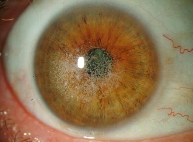
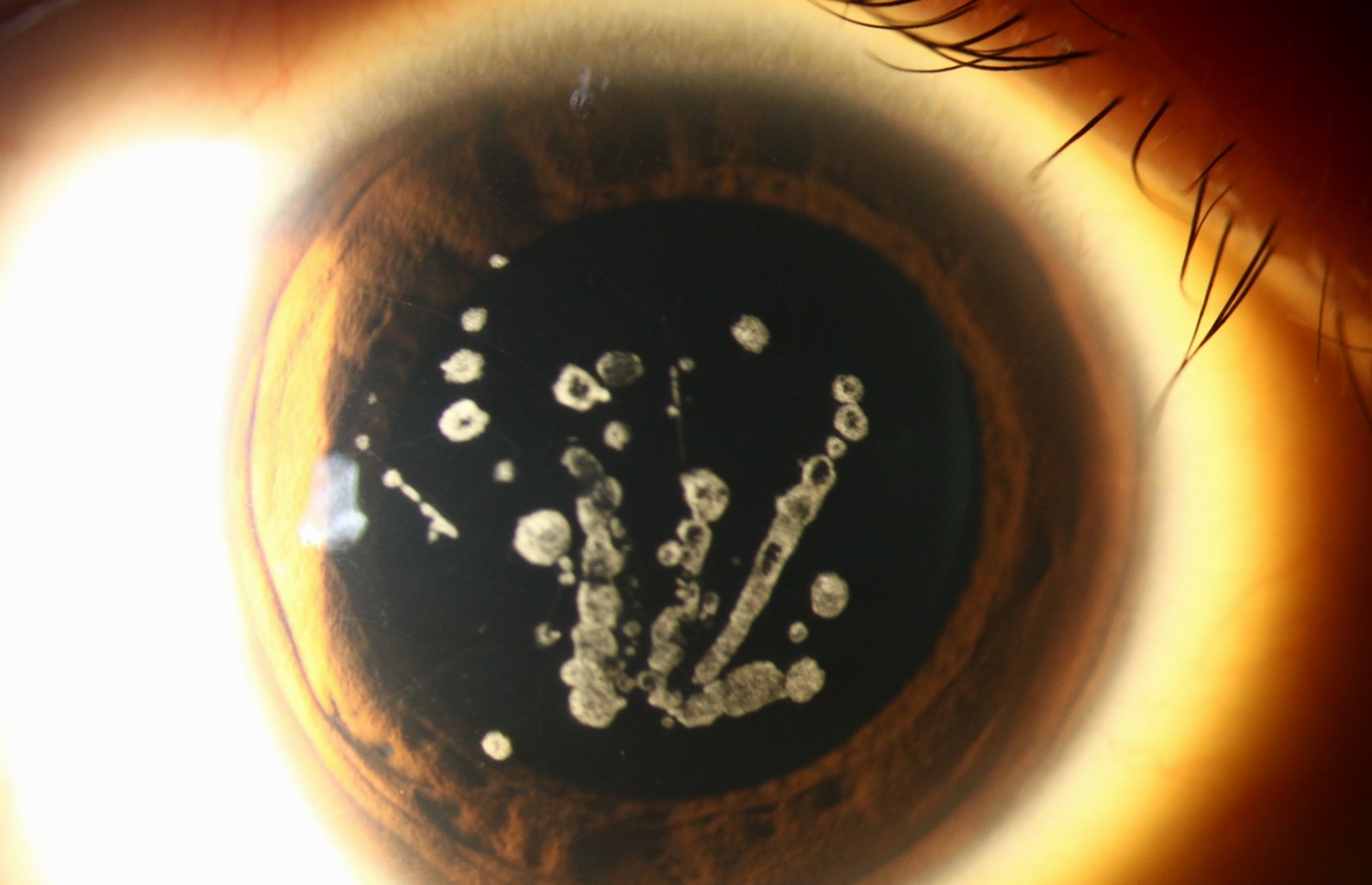
The deposits in granular corneal dystrophy are composed of hyaline material and appear as discrete, sharply demarcated, breadcrumb or snowflake-shaped white opacities scattered predominantly in the central and anterior stroma, with the surrounding stroma between deposits remaining entirely clear, a pattern often described as looking like crumbs scattered on a clean surface.
This is genetically and clinically distinct from lattice corneal dystrophy, despite both arising from mutations in the same TGFBI gene, and the clear intervening stroma is one of the most useful distinguishing features on examination.
Genetics
Granular corneal dystrophy is inherited in an autosomal dominant pattern and results from specific mutations in the TGFBI gene, distinct from the mutations that produce lattice dystrophy, and a combined granular-lattice pattern, historically called Avellino corneal dystrophy, results from yet another specific TGFBI mutation producing overlapping features of both.
All-fit Slit-Lamp Adapter
Record and share exactly what you see at the slit lamp. One adapter fits any slit lamp or surgical microscope — and any smartphone.
From Choroida — the team behind this siteClinical Course
- Onset is typically in the first decade of life, though the deposits are often small and may not be visually significant until later
- Slowly progressive, with deposits gradually increasing in number, size, and depth over decades
- Vision is often preserved reasonably well for many years, since the clear stroma between deposits allows relatively good optical transmission until the deposits become numerous and confluent enough to significantly scatter light
- Recurrent corneal erosion can occur, related to disruption of normal epithelial basement membrane adhesion over areas of deposit, though this tends to be less prominent than in lattice dystrophy
- Two clinically distinguished subtypes exist: granular corneal dystrophy type 1, with deposits generally sparing the periphery and a clear intervening stroma, and type 2 (Avellino), which combines granular and lattice-like features and tends to show somewhat different deposit characteristics and progression
Diagnosis
Diagnosis is typically made clinically based on the distinctive slit-lamp appearance of discrete white deposits with clear intervening stroma, combined with a family history consistent with autosomal dominant inheritance.
Genetic testing for the specific TGFBI mutation can confirm the diagnosis and distinguish granular dystrophy from the combined granular-lattice (Avellino) pattern, which carries somewhat different clinical implications, including a reported increased risk of unusual stromal haze following photorefractive keratectomy in affected patients.
Management
Observation
Many patients with granular corneal dystrophy have deposits that remain visually insignificant for years to decades, and observation without intervention is appropriate as long as vision remains adequate for the patient’s needs.
Phototherapeutic Keratectomy
For more superficial, anterior stromal deposits causing visual symptoms or recurrent erosion, phototherapeutic keratectomy with the excimer laser can ablate the anterior deposits and improve both surface regularity and visual clarity, offering a less invasive option than full corneal transplantation for appropriately selected patients.
Corneal Transplantation
When deposits become deep, extensive, and significantly vision-limiting, corneal transplantation, typically deep anterior lamellar keratoplasty when the endothelium is unaffected, restores clear central vision.
An Important Caution: Refractive Surgery
Patients with granular corneal dystrophy, and particularly the combined granular-lattice (Avellino) pattern, have been reported to develop unusually severe and rapid stromal haze after photorefractive keratectomy and, to a lesser extent, LASIK, so screening for a family history of corneal dystrophy before refractive surgery, and genetic testing when there is any suspicion, is an important safety step given how serious this specific complication can be.
Recurrence After Transplantation
As with other TGFBI-related stromal dystrophies, granular corneal dystrophy deposits can recur in transplanted donor tissue over years to decades, since the metabolic process producing the deposits originates from the patient’s own corneal cells rather than being eliminated by replacing central stromal tissue alone.
Prognosis
Granular corneal dystrophy generally has a favorable long-term visual prognosis given its typically slow progression and the clear stroma preserved between deposits, and both phototherapeutic keratectomy and corneal transplantation, when eventually needed, are generally effective, though with the recognized possibility of deposit recurrence over the very long term.


Document what you see
Two smartphone imaging tools built for everyday clinic use — one for the slit lamp, one for the fundus.
From Choroida — the team behind this siteReferences
- Weiss JS, Moller HU, Aldave AJ, et al. IC3D classification of corneal dystrophies–edition 2. Cornea. 2015;34:117-159.
- Rathi VM, Vyas SP, Sangwan VS. Phototherapeutic keratectomy. Br J Ophthalmol. 2012;96:618-621.
- Afshari NA, Mullally JE, Afshari MA, et al. Survey of patients with granular, lattice, avellino, and Reis-Bucklers corneal dystrophies for mutations in the BIGH3 and gelsolin genes. Arch Ophthalmol. 2001;119:16-22.
- Dinh R, Rapuano CJ, Cohen EJ, Laibson PR. Recurrence of corneal dystrophy after excimer laser phototherapeutic keratectomy. Ophthalmology. 1999;106:1490-1497.
