
Peripheral cobblestone degeneration, also called pavingstone degeneration, is one of the more common findings encountered during a thorough peripheral retinal examination, and recognizing it correctly as a low-risk, essentially benign variant, rather than a lesion carrying the same retinal detachment implications as lattice degeneration, prevents unnecessary anxiety and unnecessary prophylactic intervention.
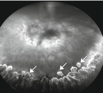
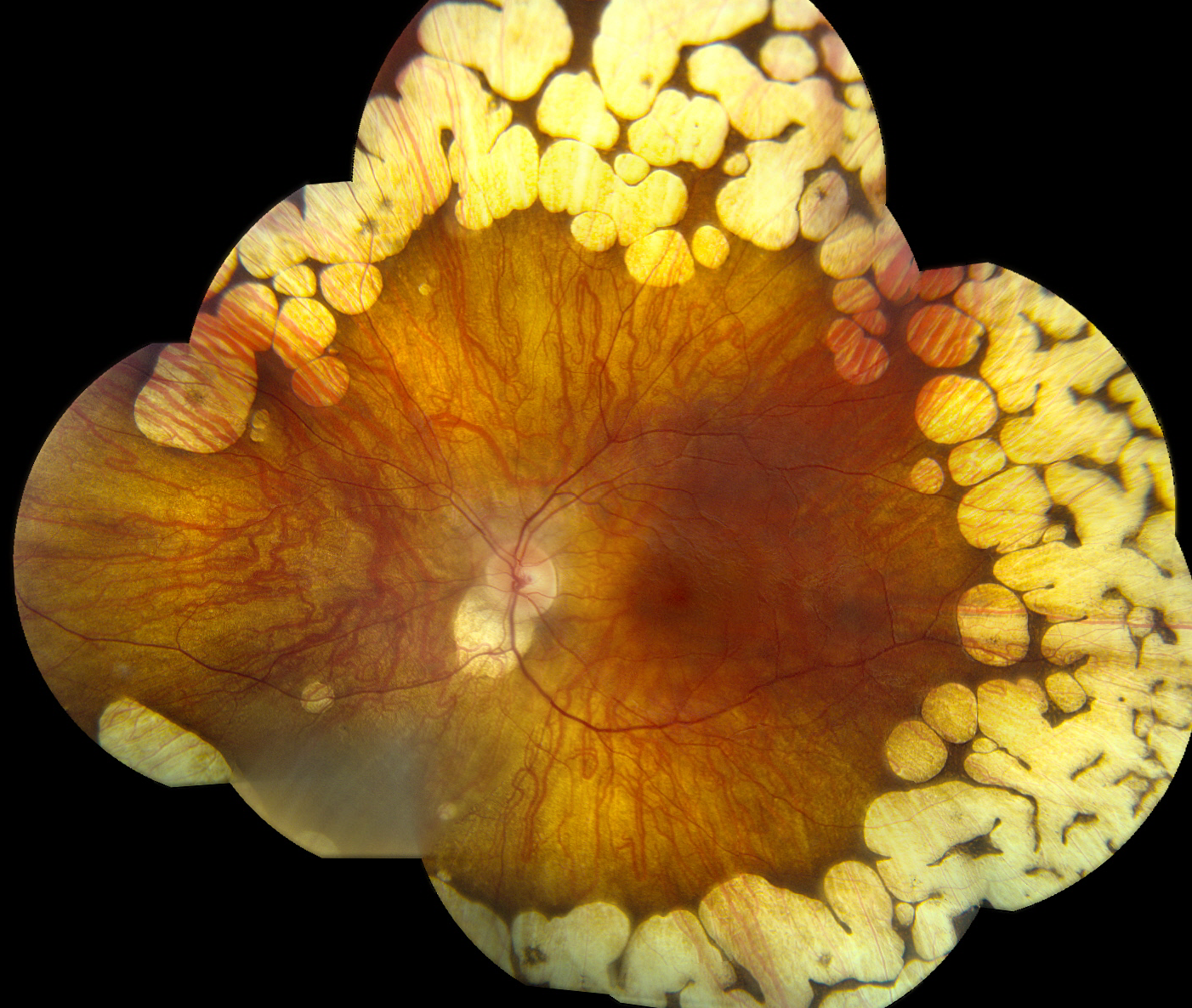
What It Looks Like
Cobblestone degeneration appears as discrete, yellow-white, sharply demarcated areas of chorioretinal atrophy, typically round to oval, often with a scalloped or cobblestone-like clustered pattern, most commonly located in the inferior peripheral retina, though it can occur in other peripheral locations as well.
Pigment clumping is frequently seen at the margins of the lesions, and choroidal vessels are often visible through the areas of retinal pigment epithelial and outer retinal atrophy, given the thinning of overlying tissue at these sites.
Why It’s Considered Low Risk
Unlike lattice degeneration, which is associated with a meaningfully increased risk of retinal tear and detachment due to areas of abnormal vitreoretinal adhesion at its margins, cobblestone degeneration does not carry this same vitreoretinal traction risk, since the lesions represent areas of chorioretinal atrophy without the abnormal, tightly adherent vitreous base characteristic of lattice degeneration.
This distinction is the central reason cobblestone degeneration is managed so differently from lattice degeneration despite both being peripheral retinal findings identified on the same dilated examination.
Fundus Explorer Pro
Photograph the retinal findings described here with the phone already in your pocket — 22 D optics and built-in illumination in one handheld unit.
From Choroida — the team behind this sitePrevalence and Associations
- A common finding, increasing in prevalence with age, and often bilateral
- Associated with myopia, though it also occurs in emmetropic and hyperopic eyes
- Generally asymptomatic and discovered incidentally during routine dilated fundus examination, particularly with scleral depression to adequately visualize the far peripheral retina where these lesions are most common
Distinguishing From Lattice Degeneration
- Cobblestone degeneration lesions are typically more discrete, rounder, and more sharply demarcated, often with visible underlying choroidal vessels, while lattice degeneration classically shows a criss-crossing pattern of sclerosed, white retinal vessels within an oval area of retinal thinning
- Lattice degeneration is more strongly associated with overlying vitreous liquefaction and firm marginal vitreoretinal adhesion, the combination that creates meaningful traction-related tear risk, a feature not characteristic of cobblestone degeneration
- Both can coexist in the same eye, and careful, complete peripheral examination with scleral depression is needed to characterize all peripheral findings accurately and distinguish between them
Clinical Evaluation
- Dilated fundus examination with scleral depression to fully visualize the far peripheral retina, since many of these lesions are located anteriorly, beyond the reach of standard non-depressed examination
- Careful description and, when feasible, wide-field fundus photography documentation, supporting comparison at future visits
- Assessment for any coexisting findings of higher-risk pathology, including lattice degeneration, retinal breaks, or areas of abnormal vitreoretinal traction, which would carry different management implications
Management
Cobblestone degeneration, identified in isolation, requires no specific treatment or prophylactic intervention, given its low associated risk of retinal tear or detachment, and periodic examination as part of routine, age-appropriate eye care is entirely sufficient.
Patients can be reassured that this specific finding does not carry the same detachment risk implications as lattice degeneration or a retinal tear, and does not require any activity restriction or prophylactic laser treatment.
When Broader Peripheral Retinal Evaluation Matters
While cobblestone degeneration itself is low-risk, its identification underscores the value of a complete, careful peripheral retinal examination in any patient undergoing dilated fundus evaluation, since this same examination is the one opportunity to identify any coexisting higher-risk findings, such as lattice degeneration or an asymptomatic retinal tear, that would change management.
Prognosis
Peripheral cobblestone degeneration has an excellent prognosis and essentially never requires treatment, remaining a stable, benign finding throughout a patient’s life in the great majority of cases.
Correctly distinguishing it from lattice degeneration and other higher-risk peripheral retinal findings is the central clinical task, allowing patients with this benign variant to be reassured while those with genuinely higher-risk findings receive the more vigilant monitoring or prophylactic treatment they need.


Document what you see
Two smartphone imaging tools built for everyday clinic use — one for the slit lamp, one for the fundus.
From Choroida — the team behind this siteReferences
- Byer NE. Long-term natural history of lattice degeneration of the retina. Ophthalmology. 1989;96:1396-1401.
- Straatsma BR, Foos RY. Typical and reticular degenerative retinoschisis. Am J Ophthalmol. 1973;75:551-575.
- Foos RY, Simons KB. Vitreous in lattice degeneration of retina. Ophthalmology. 1984;91:452-457.
- Wilkinson CP. Evidence-based analysis of prophylactic treatment of asymptomatic retinal breaks and lattice degeneration. Ophthalmology. 2000;107:12-15.
Test yourself
A few questions straight from this article.
-
Peripheral cobblestone degeneration appears on fundus examination as what?
The lesions are discrete, yellow-white, sharply demarcated areas of chorioretinal atrophy, round to oval, often clustered in a scalloped cobblestone pattern with pigment clumping at their margins. -
Where is peripheral cobblestone degeneration most commonly located?
Cobblestone degeneration sits most commonly in the inferior peripheral retina, although it can appear in other peripheral locations too. -
Why are choroidal vessels often visible within cobblestone degeneration lesions?
Retinal pigment epithelial and outer retinal atrophy thins the overlying tissue, so the choroidal vessels beneath show through the lesion. -
Why does cobblestone degeneration carry a low risk of retinal tear and detachment?
The lesions are areas of chorioretinal atrophy without the abnormal, tightly adherent vitreous base that gives lattice degeneration its traction-related tear risk. -
Which appearance identifies lattice degeneration rather than cobblestone degeneration?
Lattice degeneration classically shows a criss-crossing network of sclerosed, white retinal vessels within an oval area of retinal thinning, unlike the discrete rounded atrophy of cobblestone degeneration. -
What is the typical prevalence pattern of peripheral cobblestone degeneration?
It is a common peripheral finding whose prevalence increases with age, and it is frequently bilateral. -
Peripheral cobblestone degeneration is associated with which refractive state?
There is an association with myopia, but cobblestone degeneration is also found in emmetropic and hyperopic eyes. -
Which examination reliably detects peripheral cobblestone degeneration?
Many of these lesions lie anteriorly in the far periphery, beyond the reach of a non-depressed examination, so dilated examination with scleral depression is required. -
How is peripheral cobblestone degeneration managed when found in isolation?
Given its low tear and detachment risk, isolated cobblestone degeneration needs no prophylactic laser or activity restriction; routine age-appropriate examination is sufficient. -
What is the long-term course of peripheral cobblestone degeneration?
In the great majority of cases it remains a stable, benign finding throughout life and essentially never requires treatment.
