
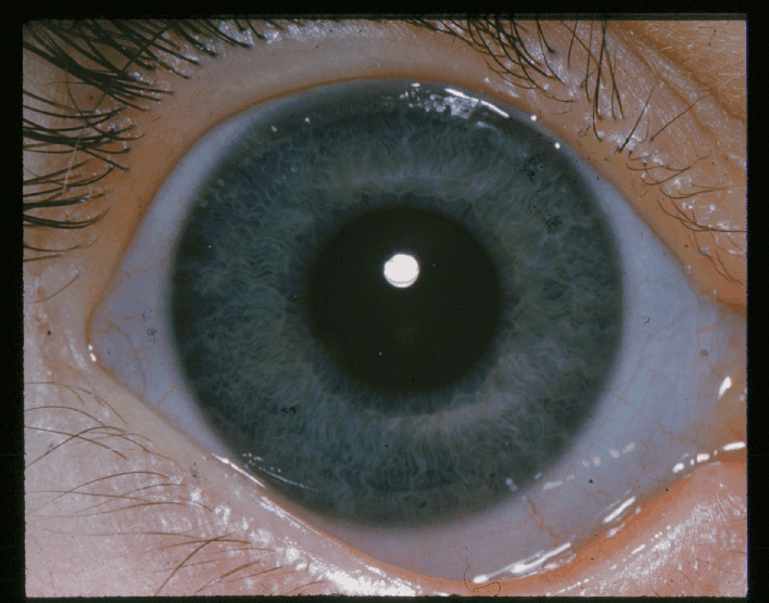
Ehlers-Danlos syndrome is a group of connective tissue disorders caused by defects in collagen synthesis or structure, and the eye, built substantially from collagen-containing tissue in the sclera and cornea, shows the underlying connective tissue fragility in ways that are often visible simply by looking closely at the eye itself.
Which subtype a given patient has matters considerably for ophthalmic risk, since the ocular findings range from a cosmetically notable but largely benign blue scleral hue in the more common subtypes to a genuine risk of spontaneous globe rupture in the rare vascular subtype, and the two should not be counselled about in the same way.
Classification and Genetics
Ehlers-Danlos syndrome comprises multiple genetically and clinically distinct subtypes, most caused by mutations affecting collagen genes or the enzymes involved in collagen processing, with the classical, hypermobile, and vascular subtypes being the most clinically relevant to ophthalmology.
The vascular subtype, caused by mutations in COL3A1 affecting type III collagen, carries by far the most serious systemic and ocular risk, including arterial and organ rupture, and is the subtype where the ocular fragility described below becomes a genuine emergency risk rather than a cosmetic finding.
Ocular Manifestations
Blue Sclera

Thinning of the sclera allows the underlying uveal pigment to show through more than usual, producing the characteristic blue-grey hue. This finding is common across several subtypes and is generally a marker of the underlying connective tissue fragility rather than a threat to vision on its own.
Corneal and Scleral Fragility
The cornea and sclera can be abnormally thin and mechanically weak, particularly in the vascular subtype, where this fragility carries a genuine risk of spontaneous or minimal-trauma rupture of the globe, a risk that shapes activity counselling and the threshold for concern after even minor ocular trauma.
Keratoconus and Corneal Ectasia
An increased prevalence of keratoconus and other ectatic corneal changes has been described in some subtypes, thought to relate to the same underlying collagen abnormality that weakens the cornea structurally, relevant to how these patients are screened before any refractive procedure.
Ocular Motility and Lid Laxity
Joint hypermobility affecting the periocular tissues can produce eyelid laxity, and some patients show mild strabismus or motility abnormalities related to the same generalised connective tissue and joint laxity seen elsewhere in the body.
Retinal Findings
Retinal detachment has been reported with somewhat increased frequency in some case series, though this association is less consistently established across all subtypes than the corneal and scleral findings.
All-fit Slit-Lamp Adapter
Record and share exactly what you see at the slit lamp. One adapter fits any slit lamp or surgical microscope — and any smartphone.
From Choroida — the team behind this siteSystemic Findings
- Joint hypermobility, ranging from mild to marked, and a central diagnostic feature across most subtypes
- Skin hyperextensibility and fragility, with easy bruising and, in some subtypes, abnormal wound healing and atrophic scarring
- Vascular fragility in the vascular subtype specifically, carrying risk of arterial dissection or rupture, and bowel or uterine rupture, making this subtype a genuinely different clinical entity from the others in terms of overall risk
Differential Diagnosis
- Osteogenesis imperfecta, which also causes blue sclera through a related but distinct collagen defect, generally distinguished by bone fragility and fracture history rather than joint hypermobility as the dominant systemic feature
- Marfan syndrome, sharing some connective tissue and lens or refractive findings but with its own distinct pattern of skeletal, cardiac, and lens (ectopia lentis) involvement rather than the scleral fragility characteristic of EDS
- Normal, physiological thin sclera in infants and some individuals without any underlying connective tissue disorder, distinguished by the absence of hypermobility, skin, or vascular findings
Diagnostic Evaluation
Diagnosis of Ehlers-Danlos syndrome relies primarily on clinical criteria specific to each subtype, incorporating joint hypermobility scoring, skin findings, and family history, with genetic testing available to confirm the molecular subtype, particularly important for distinguishing the higher-risk vascular subtype from the others.
Ophthalmic examination, including assessment of corneal thickness and topography, is useful both to characterise ocular involvement and to inform activity and safety counselling, particularly regarding contact sports or activities carrying a risk of ocular trauma in patients with the vascular subtype.
Management
There is no specific treatment for the underlying collagen defect; management is supportive and focused on monitoring and protecting at-risk tissues, coordinated with genetics and, in the vascular subtype, vascular surgery given the systemic risks involved.
Protective eyewear is advisable for patients with significant scleral or corneal fragility, particularly in the vascular subtype, given the risk of globe rupture from even relatively minor trauma that would not concern a patient with normal connective tissue.
Keratoconus, when present, is managed with standard approaches including corneal cross-linking, though decisions here should factor in the patient’s overall corneal fragility and healing capacity given the underlying connective tissue disorder.
Any ocular surgery in a patient with EDS, particularly the vascular subtype, should be planned with awareness of abnormal tissue fragility and wound healing, since standard surgical assumptions about tissue strength and healing time may not hold in the same way they do in patients without the underlying collagen defect.
Prognosis
Ocular prognosis varies considerably by subtype: blue sclera alone, seen in the more common hypermobile and classical subtypes, generally has no significant visual consequence and is best understood as a cosmetic marker of the underlying diagnosis.
In the vascular subtype, the risk of spontaneous or trauma-related globe rupture is a genuine, serious concern requiring lifelong vigilance and protective precautions, and this risk is disproportionate to how the eye may otherwise look entirely unremarkable on routine examination.
Overall prognosis is dominated by the systemic aspects of the disease, particularly vascular risk in the vascular subtype, and ophthalmic management is best understood as one component of a broader, lifelong, multidisciplinary approach to a condition that affects connective tissue throughout the body, not just the eye.


Document what you see
Two smartphone imaging tools built for everyday clinic use — one for the slit lamp, one for the fundus.
From Choroida — the team behind this siteReferences
- Malfait F, Francomano C, Byers P, et al. The 2017 international classification of the Ehlers-Danlos syndromes. American Journal of Medical Genetics Part C. 2017.
- Beighton P. Serious ophthalmological complications in the Ehlers-Danlos syndrome. British Journal of Ophthalmology. 1970.
- Segev F, Heon E, Cole WG, et al. Structural abnormalities of the cornea and lid resulting from collagen V mutations. Investigative Ophthalmology and Visual Science. 2006.
- Ehlers-Danlos Syndrome. EyeWiki, American Academy of Ophthalmology.
- Ehlers-Danlos Syndrome. StatPearls, NCBI Bookshelf.
Test yourself
A few questions straight from this article.
-
What underlies Ehlers-Danlos syndrome?
Ehlers-Danlos syndrome is a group of connective tissue disorders caused by defects in collagen synthesis or structure, which is why collagen-rich sclera and cornea show the fragility. -
What produces the blue-grey scleral hue in Ehlers-Danlos syndrome?
A thinned sclera allows the underlying uveal pigment to show through more than usual; the colour marks connective tissue fragility rather than threatening vision itself. -
Mutations in which gene define the highest-risk subtype of Ehlers-Danlos syndrome?
The vascular subtype is caused by COL3A1 mutations affecting type III collagen and carries by far the most serious systemic and ocular risk, including arterial and organ rupture. -
Which ocular risk sets the vascular subtype apart from the commoner Ehlers-Danlos subtypes?
In the vascular subtype the thin, mechanically weak cornea and sclera carry a genuine risk of globe rupture, which shapes activity counselling and the threshold for concern after minor trauma. -
Why does keratoconus matter in a patient with Ehlers-Danlos syndrome?
Keratoconus and other ectatic changes are described more often in some subtypes, thought to reflect the same collagen abnormality, which is directly relevant to refractive screening. -
Why does ocular surgery need special planning in a patient with Ehlers-Danlos syndrome?
Standard surgical assumptions about tissue strength and healing time may not hold, particularly in the vascular subtype, so operations must be planned with that fragility in mind. -
How firmly established is retinal detachment as a feature of Ehlers-Danlos syndrome?
Retinal detachment has been reported with somewhat increased frequency in some case series, but the association is less consistently established across subtypes than the corneal and scleral findings. -
What distinguishes osteogenesis imperfecta from Ehlers-Danlos syndrome when blue sclera is found?
Osteogenesis imperfecta causes blue sclera through a related but distinct collagen defect, and is distinguished by bone fragility and fractures rather than joint hypermobility as the dominant feature. -
What is advised for a patient with significant scleral or corneal fragility in Ehlers-Danlos syndrome?
Because even relatively minor trauma can rupture a fragile globe, protective eyewear is advisable, particularly in the vascular subtype, alongside counselling about contact sports. -
What dominates the overall prognosis in Ehlers-Danlos syndrome?
Ophthalmic care is one component of lifelong multidisciplinary management; overall outlook is driven by systemic disease, particularly vascular risk in the vascular subtype.

Writes and reviews the clinical articles on Educate. Every post is checked against peer-reviewed sources before it goes live.


