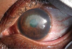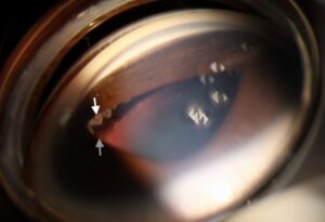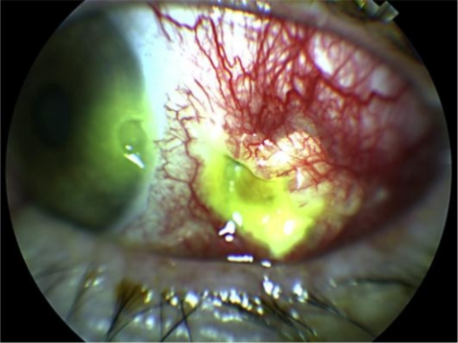
Necrotizing scleritis is the most severe and destructive form of scleral inflammation, capable of thinning the sclera to the point of perforation, and it is treated fundamentally differently from milder forms of scleritis because it so often signals an underlying systemic vasculitis that itself carries a mortality risk if left untreated.
Recognizing necrotizing disease is as much about protecting the patient’s life as protecting the eye.
Clinical Features
- Severe, often unrelenting scleral pain
- Progressive scleral thinning, sometimes visible as a bluish or grayish discoloration where underlying uvea shows through the thinned tissue
- Avascular patches of sclera, a particularly ominous sign reflecting vascular occlusion within the inflamed tissue
- Risk of scleral perforation, either spontaneously or with minimal trauma, in advanced untreated cases
- A painless variant, scleromalacia perforans, occurs almost exclusively in long-standing rheumatoid arthritis, where scleral thinning progresses with little apparent inflammation or discomfort despite similarly severe tissue destruction (see scleromalacia perforans)
Why Necrotizing Scleritis Signals Systemic Disease
Necrotizing scleritis reflects an occlusive vasculitis within the scleral and episcleral vessels, and this same vasculitic process very often affects other organs at the same time, particularly in granulomatosis with polyangiitis, where necrotizing scleritis can be a presenting or defining feature of active systemic disease.
Historical data on necrotizing scleritis associated with systemic vasculitis showed a substantial risk of death within a few years when the underlying vasculitis was inadequately treated, a finding that fundamentally shaped current management, which prioritizes systemic immunosuppression over local eye treatment alone.
All-fit Slit-Lamp Adapter
Record and share exactly what you see at the slit lamp. One adapter fits any slit lamp or surgical microscope — and any smartphone.
From Choroida — the team behind this siteWorkup
- Full systemic vasculitis screen: ANCA (both c-ANCA/PR3 and p-ANCA/MPO), rheumatoid factor and anti-CCP antibodies, ANA, complement levels, and inflammatory markers
- Renal function testing and urinalysis, since renal involvement is common in ANCA-associated vasculitis and may be asymptomatic
- Chest imaging, looking for pulmonary nodules or infiltrates that can accompany granulomatosis with polyangiitis
- Infectious workup, including syphilis and tuberculosis testing, and specific consideration of infectious causes after any recent ocular surgery, since infectious scleritis needs antimicrobial rather than immunosuppressive treatment
- Coordination with rheumatology from the point of diagnosis, given the frequency and seriousness of systemic association
Management
Systemic Corticosteroids
High-dose systemic corticosteroids are used to gain initial control of the inflammation, but they are not considered adequate as sole long-term therapy for necrotizing scleritis associated with systemic vasculitis.
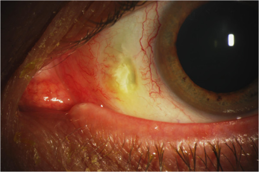
Steroid-Sparing Immunosuppression
Cyclophosphamide has historically been the standard agent for severe, vasculitis-associated necrotizing scleritis, particularly with granulomatosis with polyangiitis, given its established role in treating the systemic disease.
Rituximab has become an important alternative, with strong evidence in ANCA-associated vasculitis generally and increasing use specifically for ocular manifestations including necrotizing scleritis, often preferred now over cyclophosphamide given a more favorable side-effect profile for many patients.
Other agents, including methotrexate and mycophenolate, are used for less severe or non-vasculitic necrotizing disease.
Topical and Local Measures
Topical treatment plays a supportive role at best in true necrotizing scleritis, since the disease is fundamentally systemic; topical or periocular corticosteroid injections are generally avoided or used cautiously given a theoretical risk of worsening scleral thinning.
Surgical Management
Impending or actual scleral perforation may require surgical reinforcement with a scleral, corneal, or periosteal patch graft, performed alongside, not instead of, systemic immunosuppression, since surgery alone does not address the underlying inflammatory process driving further thinning.
Prognosis
With prompt recognition and appropriate systemic immunosuppression, both ocular and systemic outcomes have improved substantially compared with the era before modern immunosuppressive and biologic therapy.
Delayed diagnosis or inadequate systemic treatment carries real risk, both of scleral perforation and vision loss, and of the broader morbidity and mortality associated with untreated systemic vasculitis, which is the central reason necrotizing scleritis is managed as a systemic disease with an ocular manifestation rather than as an eye disease alone.


Document what you see
Two smartphone imaging tools built for everyday clinic use — one for the slit lamp, one for the fundus.
From Choroida — the team behind this siteReferences
- Sainz de la Maza M, Foster CS, Jabbur NS. Scleritis associated with rheumatoid arthritis and with other systemic immune-mediated diseases. Ophthalmology. 1994;101:1281-1286.
- Foster CS, Forstot SL, Wilson LA. Mortality rate in rheumatoid arthritis patients developing necrotizing scleritis or peripheral ulcerative keratitis. Ophthalmology. 1984;91:1253-1263.
- Ahn SJ, Kim NR, Kim CY, Lee ES. Efficacy and safety of rituximab in refractory noninfectious scleritis. Ocul Immunol Inflamm. 2021;29:369-377.
- Stone JH, Merkel PA, Spiera R, et al. Rituximab versus cyclophosphamide for ANCA-associated vasculitis. N Engl J Med. 2010;363:221-232.
Test yourself
A few questions straight from this article.
-
Necrotizing scleritis reflects which underlying vascular process?
Necrotizing scleritis reflects an occlusive vasculitis within the scleral and episcleral vessels, and the same vasculitic process very often involves other organs simultaneously. -
In necrotizing scleritis, avascular patches of sclera are an ominous sign of what?
Avascular patches of sclera are described as a particularly ominous sign because they reflect vascular occlusion within the inflamed tissue. -
A bluish or grayish scleral discoloration in necrotizing scleritis represents what?
Progressive scleral thinning may become visible as a bluish or grayish discoloration where the underlying uvea shows through the thinned tissue. -
Necrotizing scleritis can be a presenting or defining feature of which systemic vasculitis?
In granulomatosis with polyangiitis, necrotizing scleritis can be a presenting or defining feature of active systemic disease. -
What is the role of high-dose systemic corticosteroids in vasculitis-associated necrotizing scleritis?
High-dose systemic corticosteroids gain initial control of the inflammation but are not adequate as sole long-term therapy when systemic vasculitis underlies the disease. -
Which agent has historically been standard for severe vasculitis-associated necrotizing scleritis?
Cyclophosphamide has historically been the standard agent for severe vasculitis-associated necrotizing scleritis, particularly with granulomatosis with polyangiitis, given its established role in the systemic disease. -
Why is rituximab often now preferred over cyclophosphamide in necrotizing scleritis?
Rituximab has strong evidence in ANCA-associated vasculitis and is often preferred over cyclophosphamide because its side-effect profile is more favorable for many patients. -
Why do renal function tests and urinalysis belong in the necrotizing scleritis workup?
Renal function testing and urinalysis are needed because renal involvement is common in ANCA-associated vasculitis and may be asymptomatic. -
Why are periocular corticosteroid injections generally avoided in necrotizing scleritis?
Topical measures are supportive at best, and topical or periocular corticosteroid injections are avoided or used cautiously given a theoretical risk of worsening scleral thinning. -
How should a patch graft for impending scleral perforation be used?
Scleral, corneal or periosteal patch grafting is performed alongside systemic immunosuppression, because surgery alone does not address the inflammatory process driving further thinning.

Writes and reviews the clinical articles on Educate. Every post is checked against peer-reviewed sources before it goes live.