Cup to disc ratio is the size of the optic cup divided by the size of the optic disc, and it is a basic measure in glaucoma assessment. Most healthy eyes measure about 0.3 to 0.4 vertically. Because the number depends on disc size, read it together with the rim, the disc size and the other eye.
What Is Cup to Disc Ratio?
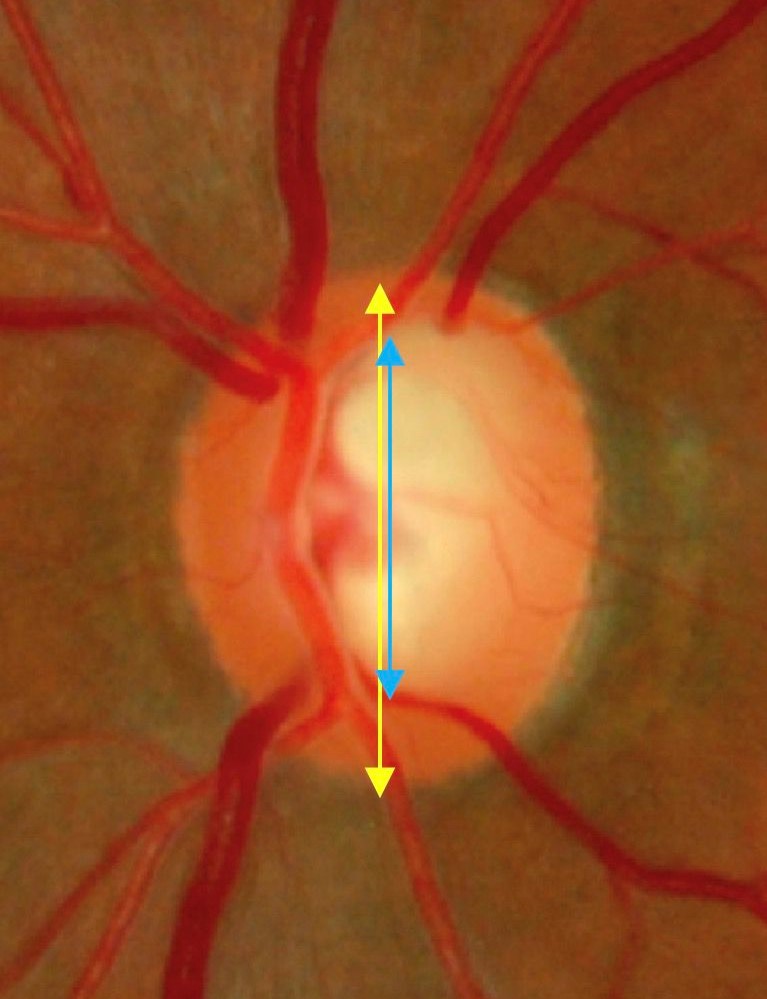
The cup to disc ratio compares the width of the central cup with the width of the whole disc, and it runs from 0.0 to 1.0. Clinicians usually record the vertical ratio, because glaucoma tends to elongate the cup vertically. The terms cup-to-disc, cup disk ratio, disc cup ratio and optic disc ratio all describe the same measurement.
Cup to Disc Ratio in the Eye: Cup, Rim and Disc
The disc is the whole optic nerve head, where about a million retinal ganglion cell axons leave the eye. The cup is the pale central depression with no nerve fibers, and the neuroretinal rim is the pink tissue between the cup and the disc edge. The rim carries the fibers, so it matters most.
Fundus Explorer Pro
Photograph the retinal findings described here with the phone already in your pocket — 22 D optics and built-in illumination in one handheld unit.
From Choroida — the team behind this siteNormal Cup to Disc Ratio
Most healthy eyes have a vertical ratio between 0.3 and 0.4, and the upper limit of normal is about 0.6 in most populations. Armaly showed that cup size is largely inherited, so a patient with a large cup may come from a family with large cups and no disease. Check the family history before you call a large cup abnormal.
What Cup to Disc Ratio Is Concerning?
A vertical ratio above 0.6, a ratio that rises between visits, or a ratio much larger than the other eye is concerning. A ratio of 0.5 is common in healthy eyes and needs no action alone. Think about disc size, rim shape and the patient’s risk factors, such as high pressure, age and family history of glaucoma.
Disc Size and the Optic Cup to Disc Ratio
Large discs have larger cups and small discs have smaller cups, so a ratio of 0.6 can be normal in a large disc and abnormal in a small one. Garway-Heath and colleagues showed that relating the ratio to disc size improves the assessment. Because disc size varies a lot, judge it first and then judge the cup against it.
Magnification also changes the apparent disc size. A 90 diopter lens shows a smaller image than a 78 diopter lens, and a direct ophthalmoscope magnifies the disc more, so correct for the lens you use when you size the disc.
Cup to Disc Ratio in Glaucoma
In glaucoma, loss of nerve fibers thins the rim, and the cup enlarges as the rim shrinks. A large ratio raises suspicion, but no single value diagnoses glaucoma. Confirm it with intraocular pressure measurement, visual fields and imaging.
Besides a large ratio, look for these rim signs of glaucoma:
- Focal notching or thinning of the rim, most often inferior or superior
- Baring of circumlinear vessels and bayoneting and nasalization of vessels
- Retinal nerve fiber layer defects that match the rim damage
- Progressive change in the rim between visits
Rim Assessment and the ISNT Rule
The rim shape matters more than the ratio. The ISNT rule describes the normal pattern, with the rim thickest inferiorly, then superiorly, nasally and temporally. Many healthy eyes break the rule, so treat it as a prompt and examine every quadrant for thinning or notching.
How to Examine the Optic Disc
Dilate the pupil and use a slit lamp with a handheld 78 or 90 diopter lens, because a stereoscopic view shows the contour of the cup and the rim. The direct ophthalmoscope gives a magnified but monocular view. Record the cup and disc sizes in each eye, and note any notching or hemorrhage (see optic disc hemorrhage).
Judge the cup edge by the bend of the vessels as they cross the rim, not by the pallor alone, because the pale area can be smaller than the true cup. Use a narrow beam, a steady lens and a fine focus to see the contour.
Asymmetry Between the Eyes
A difference of more than 0.2 between the eyes raises suspicion, but asymmetry can be physiologic when the discs differ in size. Compare the rim and the visual field before you diagnose disease. Asymmetry is a trigger for more testing, not a diagnosis.
Where the Cup to Disc Ratio Misleads
Observers differ a lot when they estimate the ratio, and tilted discs and myopic discs distort the apparent cup (see tilted disc). Because glaucoma can damage the rim without enlarging a small cup much, follow the rim and photographs instead of a single number.
The ratio does not capture cup depth, the rim area or the retinal nerve fiber layer thickness. Two eyes with the same ratio can have very different rim areas when the discs differ in size, so look at the rim directly and use imaging to measure it.
Other disc conditions also change the apparent cup. A dragged disc distorts the contour, and buried optic disc drusen can crowd a small disc and mimic swelling. Measure the ratio only when the disc outline is clear.
Cupping Versus Pallor
Glaucoma produces cupping with loss of the rim, while other optic neuropathies produce pallor with little cupping (see optic atrophy). Compressive and ischemic lesions can also produce cupping in some patients, so look at the color of the rim as well as its shape. Pallor out of proportion to the cup argues against glaucoma.
Cup to Disc Ratio in Children
Large cups in children may reflect high pressure or congenital glaucoma, and cupping can partly reverse after pressure control. Physiologic cups in children are small, so take an enlarged cup in a child seriously and check pressure and corneal findings.
Race and Age
Average disc and cup size varies with ethnicity, and people of African descent have larger discs and larger cups on average. Aging does not change cup size much in healthy eyes, so a cup that grows over time deserves attention. Apply population norms with care.
Imaging and Follow-Up
Disc photographs give a permanent record for comparison, and stereo photographs show the contour better. Optical coherence tomography adds objective measures of the rim and the retinal nerve fiber layer. Store images at baseline and at regular intervals, because progression matters more than any single reading (see primary open-angle glaucoma).
Explaining the Result to Patients
Explain the ratio in plain terms and say that it varies from person to person. Avoid labeling a patient with a large cup as having glaucoma without more evidence, and explain the plan for follow-up and tests. A large cup with a healthy rim, normal pressure and normal fields is often a physiologic variant.
Practical Tips at the Slit Lamp
Compare both eyes in the same session with the same lens. Practice grading a few photographs with a colleague, because small details change the estimate and agreement between observers is modest. Write the ratio, the disc size and the rim description in the chart each time.


Document what you see
Two smartphone imaging tools built for everyday clinic use — one for the slit lamp, one for the fundus.
From Choroida — the team behind this siteReferences
- Armaly MF. Genetic determination of cup/disc ratio of the optic nerve. Arch Ophthalmol. 1967;78:35-43.
- Lichter PR. Variability of expert observers in evaluating the optic disc. Trans Am Ophthalmol Soc. 1976;74:532-572.
- Garway-Heath DF, Ruben ST, Viswanathan A, Hitchings RA. Vertical cup/disc ratio in relation to optic disc size: its value in the assessment of the glaucoma suspect. Br J Ophthalmol. 1998;82:1118-1124.
- Weinreb RN, Aung T, Medeiros FA. The pathophysiology and treatment of glaucoma: a review. JAMA. 2014;311:1901-1911.

Writes and reviews the clinical articles on Educate. Every post is checked against peer-reviewed sources before it goes live.