
A pale optic disc is not a diagnosis. It is the scar left behind by one, and the real work is finding out which.
Optic atrophy is the visible end point of irreversible loss of retinal ganglion cell axons, whatever destroyed them.
By the time the disc turns pale, the damage that caused it has usually already finished.
That makes optic atrophy a fundamentally backward-looking sign – and a forward-looking warning about the fellow eye and the underlying cause.
Some causes are benign and burnt out; others are compressive, hereditary or nutritional and demand urgent action to protect what remains.
Reading the pattern of pallor, and pairing it with the history and the fellow eye, is what turns a descriptive finding into an actionable diagnosis.
What Is Optic Atrophy?
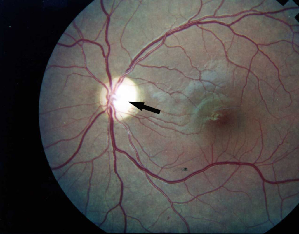
Optic atrophy is pallor of the optic disc resulting from permanent loss of retinal ganglion cell axons and their supporting glial and vascular tissue.
It is described by the pattern of pallor and by the mechanism behind it:
- Primary optic atrophy – a chalky white, sharply defined disc with no preceding swelling, typical of compressive, hereditary or toxic causes
- Secondary optic atrophy – a greyish, indistinct disc with blurred margins, following resolved papilledema or optic neuritis
- Consecutive optic atrophy – waxy pallor following primary retinal disease such as retinitis pigmentosa
- Glaucomatous optic atrophy – pallor accompanied by pathological cupping, a distinct pattern from all of the above
The distinction matters clinically because the pattern of pallor points back toward the category of disease that produced it.
Epidemiology
Optic atrophy is a common end point rather than a single disease, so its epidemiology is really that of its causes.
- Glaucoma is the single largest contributor worldwide when glaucomatous optic neuropathy is included
- Ischaemic optic neuropathy is the commonest non-glaucomatous cause in older adults
- Inflammatory and demyelinating causes predominate in younger adults
- Hereditary optic neuropathies such as Leber hereditary optic neuropathy and dominant optic atrophy typically present in childhood or early adulthood
- Compressive lesions can occur at any age, and are the causes with the most to gain from early detection
Because the age of the patient shifts the likely cause so substantially, age is one of the most useful early filters when working up a pale disc.
Fundus Explorer Pro
Photograph the retinal findings described here with the phone already in your pocket — 22 D optics and built-in illumination in one handheld unit.
From Choroida — the team behind this sitePathophysiology
The pale appearance has a specific structural basis.
- An insult – ischaemic, inflammatory, compressive, toxic, hereditary or traumatic – injures retinal ganglion cell axons
- Axonal degeneration proceeds along the visual pathway, and the ganglion cell bodies are lost
- The retinal nerve fibre layer thins, most readily measured on OCT
- Loss of axons is accompanied by loss of the fine capillary network on the disc surface, which is a major contributor to the pallor
- Glial tissue replaces the lost axons, producing the final atrophic appearance
The clinically important consequence is that this process is not reversible – treatment is aimed at the underlying cause and at protecting surviving axons, never at restoring lost ones.
Causes and Risk Factors
Acquired Causes
- Glaucoma, producing pallor with characteristic cupping
- Ischaemic optic neuropathy, both arteritic and non-arteritic
- Optic neuritis and other inflammatory optic neuropathies
- Compressive lesions – pituitary adenoma, meningioma, aneurysm and orbital masses
- Traumatic optic neuropathy
- Chronic papilledema progressing to secondary atrophy
Hereditary, Nutritional and Toxic Causes
- Leber hereditary optic neuropathy and autosomal dominant optic atrophy
- Vitamin B12 and folate deficiency
- Toxic exposures including methanol, ethambutol, amiodarone and tobacco-alcohol amblyopia
- Syndromic associations such as Wolfram syndrome
The nutritional and toxic causes deserve particular attention because they are among the few in which further loss is genuinely preventable by intervention.
Clinical Presentation
Symptoms
- Reduced visual acuity, ranging from mild to severe depending on the extent of axonal loss
- Loss of colour vision, characteristically disproportionate to the acuity, with red desaturation an early sign
- Reduced contrast sensitivity and a subjective sense that vision is “dim” or “washed out”
- Visual field defects whose pattern reflects the site of the original insult
- A history that may be silent, since gradual bilateral loss is easily missed by the patient
Examination Findings
- Pallor of the optic disc, ranging from subtle temporal pallor to a chalky white disc
- Loss of the fine capillary network normally visible on the disc surface
- A relative afferent pupillary defect in unilateral or markedly asymmetric disease
- Thinning of the retinal nerve fibre layer, sometimes visible clinically and reliably measured on OCT
- Reduced colour vision on Ishihara or a formal colour test, often out of proportion to the Snellen acuity
- Cupping alongside pallor, which points specifically toward a glaucomatous mechanism
Pallor confined to the temporal disc is easy to overlook and often the earliest visible sign, particularly in toxic, nutritional and hereditary optic neuropathies.
Diagnostic Evaluation
Structural and Functional Assessment
- OCT of the retinal nerve fibre layer and ganglion cell complex to quantify axonal loss objectively
- Formal automated perimetry, since the field defect pattern localises the lesion along the visual pathway
- Colour vision testing and assessment for a relative afferent pupillary defect
Neuroimaging
- MRI of the brain and orbits with contrast to exclude a compressive or infiltrative lesion, which is the single most important thing not to miss
- Imaging is particularly urgent in unexplained unilateral atrophy, in progressive loss, and in any patient with a junctional or bitemporal field defect
Laboratory and Genetic Testing
- Vitamin B12, folate and, where relevant, thiamine levels
- Inflammatory and infectious serology when the history suggests it
- ESR and CRP where arteritic ischaemic optic neuropathy is a consideration
- Genetic testing for hereditary optic neuropathies in young patients, bilateral disease or a suggestive family history
An unexplained pale disc without imaging is an incomplete assessment – a compressive lesion is the diagnosis whose delay costs the most.
Differential Diagnosis
Findings that can be mistaken for true optic atrophy include:
- A physiologically pale but healthy disc, particularly a large disc with a big cup
- Optic disc hypoplasia – a small disc, present from birth, with the double-ring sign rather than acquired pallor
- Myelinated retinal nerve fibres, whose white appearance can be mistaken for pallor
- Optic disc drusen, which distort the disc appearance and can coexist with genuine field loss
- Glaucomatous optic neuropathy, which is a specific pattern rather than a mimic, and is distinguished by the cupping-to-pallor relationship
The most reliable way to separate genuine atrophy from a pale-looking normal disc is to pair the appearance with objective evidence: OCT thinning, a field defect, reduced colour vision or a relative afferent pupillary defect.
Management
Treating the Underlying Cause
- Urgent decompression of a compressive lesion, which can preserve and sometimes recover function in surviving axons
- High-dose corticosteroids and urgent systemic treatment where giant cell arteritis is suspected, primarily to protect the fellow eye
- Correction of vitamin B12, folate or thiamine deficiency
- Withdrawal of an implicated toxic agent such as ethambutol
- Intraocular pressure reduction where the mechanism is glaucomatous
Protecting the Fellow Eye
- Many causes are bilateral or sequential, so the fellow eye is often the real target of treatment
- Genetic counselling where a hereditary optic neuropathy is confirmed, including implications for relatives
Visual Rehabilitation
- Low vision assessment, magnification aids and lighting optimisation
- Registration for visual impairment support where appropriate
- Counselling regarding driving and occupational implications
Because the atrophy itself cannot be reversed, the entire therapeutic effort is directed at the cause, the fellow eye and the patient’s remaining function.
Prognosis
Visual prognosis depends almost entirely on the underlying cause and on how much axonal reserve remains.
- Established pallor represents permanent axonal loss and does not recover
- Some function can improve if a compressive lesion is decompressed before axons are lost, which is why timing dominates outcome
- Progressive causes – hereditary, toxic, nutritional and glaucomatous – can continue to worsen unless the underlying process is addressed
- Bilateral involvement is common in hereditary, nutritional and toxic disease, so a unilateral presentation often does not stay unilateral
The practical message is that a pale disc should never be filed as an endpoint: it is a prompt to identify the cause and to protect whatever vision, in either eye, is still salvageable.


Document what you see
Two smartphone imaging tools built for everyday clinic use — one for the slit lamp, one for the fundus.
From Choroida — the team behind this siteReferences
- Miller NR, Newman NJ, Biousse V, Kerrison JB. Walsh and Hoyt’s Clinical Neuro-Ophthalmology.
- Yu-Wai-Man P, Griffiths PG, Chinnery PF. Mitochondrial optic neuropathies – disease mechanisms and therapeutic strategies. Progress in Retinal and Eye Research. 2011.
- Behbehani R. Clinical approach to optic neuropathies. Clinical Ophthalmology. 2007.
- Optic Atrophy. StatPearls, NCBI Bookshelf.
- Optic Atrophy. EyeWiki, American Academy of Ophthalmology.
Test yourself
A few questions straight from this article.
-
What does optic disc pallor in optic atrophy actually represent?
Optic atrophy is pallor resulting from permanent loss of retinal ganglion cell axons together with their supporting glial and vascular tissue, so it is the scar of a finished insult. -
A chalky white, sharply defined disc with no preceding swelling describes which pattern of optic atrophy?
Primary optic atrophy is a chalky white, sharply defined disc that was never swollen, and it is typical of compressive, hereditary or toxic causes. -
Which structural change is a major contributor to the pale appearance of the atrophic disc?
Axonal loss is accompanied by loss of the fine capillary network on the disc surface, a major contributor to the pallor, with glial tissue replacing the lost axons. -
Which is the commonest non-glaucomatous cause of optic atrophy in older adults?
Ischaemic optic neuropathy is the commonest non-glaucomatous cause in older adults, while inflammatory and demyelinating causes predominate in younger adults. -
Which sensory finding is characteristically disproportionate to the acuity in optic atrophy?
Colour vision loss is characteristically out of proportion to the Snellen acuity, with red desaturation an early sign, alongside reduced contrast sensitivity. -
Which disc appearance is often the earliest visible sign in toxic, nutritional and hereditary optic neuropathies?
Temporal pallor is easy to overlook and is often the earliest visible sign, particularly in toxic, nutritional and hereditary optic neuropathies. -
Which investigation addresses the diagnosis that is most important not to miss in unexplained optic atrophy?
MRI of the brain and orbits with contrast excludes a compressive or infiltrative lesion, the single most important thing not to miss, and is especially urgent in unexplained unilateral or progressive atrophy. -
Why are high-dose corticosteroids given urgently when giant cell arteritis is suspected?
High-dose corticosteroids and urgent systemic treatment in suspected giant cell arteritis are given primarily to protect the fellow eye, since established atrophy cannot be reversed. -
Which feature identifies optic disc hypoplasia rather than acquired optic atrophy?
Optic disc hypoplasia is a small disc present from birth showing the double-ring sign, rather than the acquired pallor of true optic atrophy. -
What determines whether any visual function can be regained in compressive optic atrophy?
Established pallor represents permanent axonal loss and does not recover, but some function can improve if a compressive lesion is decompressed before axons are lost, so timing dominates outcome.

Writes and reviews the clinical articles on Educate. Every post is checked against peer-reviewed sources before it goes live.


