Vision lost to glaucoma is, in principle, preventable — but only if the disease is caught early enough to act.
Primary open-angle glaucoma (POAG) is the condition that tests this promise more than any other.
It is the most common form of glaucoma worldwide and a leading cause of irreversible blindness.
Yet it rarely announces itself.
Intraocular pressure (IOP) can be high, borderline, or entirely normal at the time of diagnosis, and the earliest visual field loss sits outside the area a patient actually looks at.
By the time central vision is affected, the optic nerve has often already lost a substantial fraction of its retinal ganglion cell axons.
Understanding how POAG damages the optic nerve, why it hides so effectively from patients and routine screening alike, and how to catch it early is essential knowledge for every eye-care professional.
What Is Primary Open-Angle Glaucoma?
Primary open-angle glaucoma (POAG) is a chronic, progressive optic neuropathy defined by a triad of structural and functional findings, not by a pressure number alone.
The diagnosis requires:
- An open, normal-appearing anterior chamber angle on gonioscopy
- Progressive loss of retinal ganglion cells and their axons
- Characteristic cupping and thinning of the optic nerve head
- Corresponding visual field loss, typically peripheral before central
- Elevated intraocular pressure in most, but not all, affected eyes
POAG is ultimately a disease of the optic nerve — the angle is open and the eye looks otherwise unremarkable, which is precisely why it is so easy to overlook.
Epidemiology
POAG is the most common subtype of glaucoma and one of the leading causes of irreversible vision loss globally.
Key figures from large-scale meta-analysis:
- Worldwide age-standardized prevalence of glaucoma in people aged 40 and older is approximately 3.5%
- Global glaucoma cases were estimated at 52.7 million in 2020, projected to rise to roughly 79.8 million by 2040
- The burden falls disproportionately on African and Asian populations as these populations age
- People of African ancestry develop POAG more frequently, often earlier, and with a higher risk of blindness than people of European ancestry
- A first-degree relative with POAG substantially increases an individual’s own risk
As the global population ages, the absolute number of people living with undiagnosed POAG is expected to keep climbing.
Fundus Explorer Pro
Photograph the retinal findings described here with the phone already in your pocket — 22 D optics and built-in illumination in one handheld unit.
From Choroida — the team behind this sitePathophysiology
Despite an open angle, aqueous outflow through the trabecular meshwork becomes progressively less efficient in POAG.
Extracellular matrix changes and increased outflow resistance at the level of the trabecular meshwork and juxtacanalicular tissue raise intraocular pressure, even though the angle remains anatomically open on gonioscopy.
This chain of events is thought to unfold roughly as follows:
- Elevated IOP mechanically stresses the lamina cribrosa at the optic nerve head
- Axonal transport within retinal ganglion cell axons is disrupted at this point of stress
- Impaired optic nerve head blood flow and vascular dysregulation compound the mechanical injury in many patients
- Retinal ganglion cells undergo progressive apoptosis
- Axonal loss produces retinal nerve fiber layer (RNFL) thinning
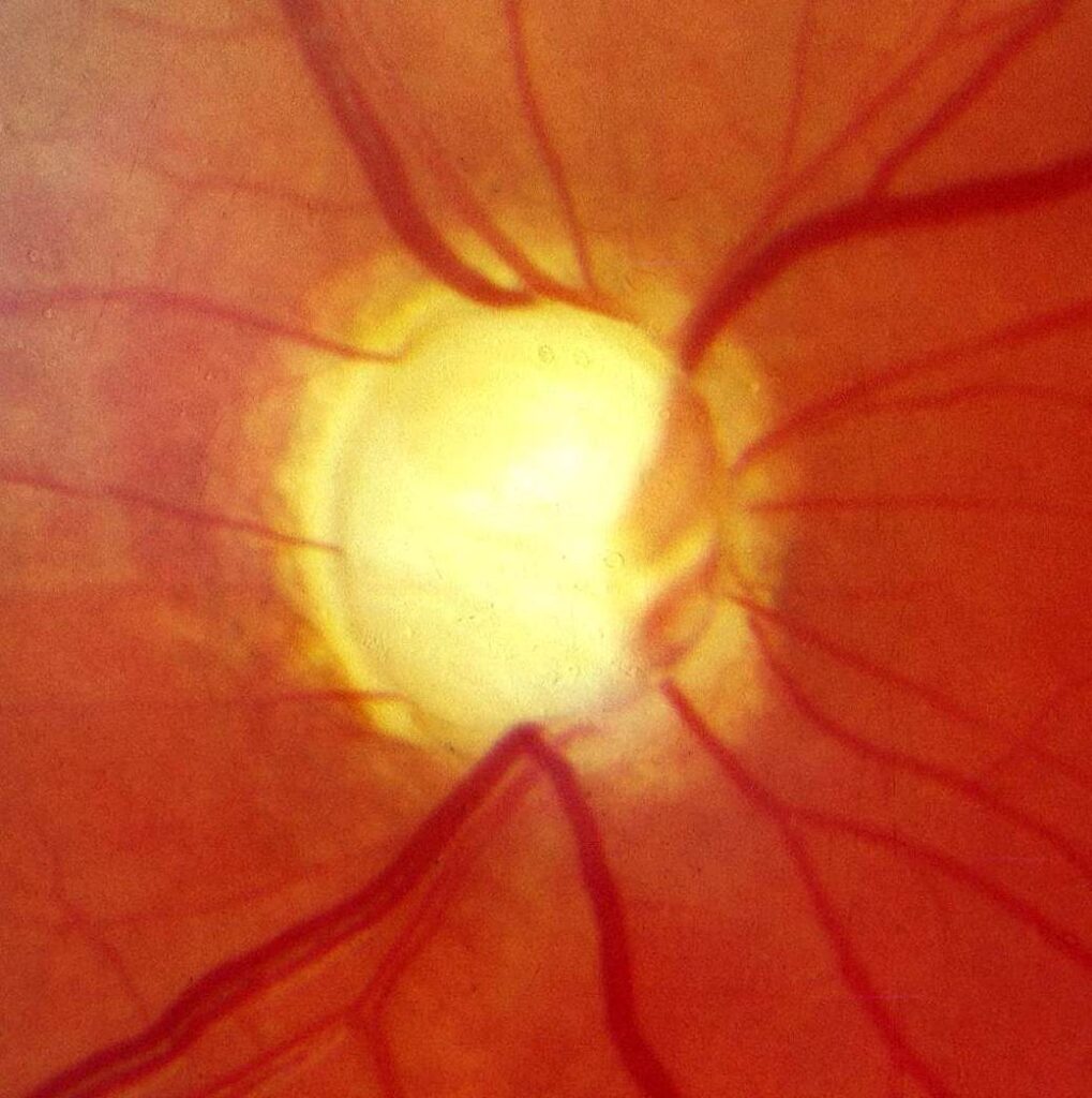
- Loss of the neuroretinal rim produces the clinically visible cupping of the optic disc
Both the mechanical and vascular mechanisms are supported by evidence, and most cases likely involve a combination of the two rather than either factor alone.
Risk Factors
Ocular Risk Factors
- Elevated intraocular pressure — the single strongest and only modifiable risk factor
- Thin central corneal thickness, independently associated with progression to POAG in ocular hypertensive eyes
- Myopia, particularly moderate to high myopia
- Optic disc hemorrhage at baseline
- A larger vertical cup-to-disc ratio at initial examination
Demographic and Systemic Risk Factors
- Increasing age
- Family history of glaucoma in a first-degree relative
- African or Hispanic ancestry
- Possible association with vascular dysregulation (migraine, Raynaud phenomenon) — evidence remains debated
- Diabetes mellitus, though the strength of this association is inconsistent across studies
Elevated IOP remains the only risk factor clinicians can directly modify, which is why it anchors both diagnosis and treatment.
Clinical Presentation
Symptoms
POAG is classically asymptomatic through most of its course.
Peripheral visual field loss progresses so gradually that patients rarely notice it until it is advanced, since the fellow eye and normal cortical compensation mask early deficits.
Only in later stages do patients report:
- Difficulty with night driving
- Missing steps or bumping into objects on one side
- A subjective sense of “tunnel vision”
Central visual acuity is typically preserved until disease is quite advanced — which is exactly why relying on a patient’s own vision complaints delays diagnosis.
Examination Findings
The optic nerve head carries the key clinical evidence. Findings on dilated fundus examination include:
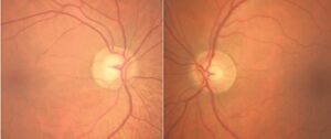
- An increased or asymmetric cup-to-disc ratio between the two eyes
- Focal thinning or notching of the neuroretinal rim, often at the inferior or superior poles
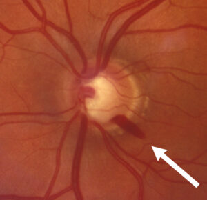
- Splinter-shaped disc hemorrhages, a marker of active progression
- Wedge-shaped retinal nerve fiber layer defects on red-free photography
- An open angle on gonioscopy, visible in all four quadrants
These structural changes routinely precede any visual field abnormality that the patient can detect — which is exactly why POAG has earned the nickname “the silent thief of sight.”
Why Primary Open-Angle Glaucoma Is So Often Missed
Several features of POAG conspire against early detection.
- There are no early symptoms to prompt the patient to seek care
- IOP alone is an unreliable screening tool — a meaningful proportion of POAG occurs with pressures inside the statistically normal range (normal-tension glaucoma), particularly in Asian populations
- Automated visual field testing is not part of routine general eye or medical check-ups
- Early field defects are typically arcuate or paracentral, sparing central fixation and going unnoticed by the patient
- A physiologically large but stable cup-to-disc ratio can resemble pathological cupping without baseline photographs or OCT for comparison
Because none of these gaps closes on its own, a deliberate, structured evaluation — not a passing pressure check — is what actually catches the disease early.
Diagnostic Evaluation
Tonometry
Goldmann applanation tonometry remains the reference standard for measuring IOP.
- IOP shows diurnal variation, so a single reading can miss peak pressures
- Measured IOP should be interpreted alongside central corneal thickness, since thin corneas can underestimate true pressure
Gonioscopy
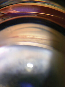
Gonioscopy is mandatory, not optional, in every suspected case.
- Confirms an open angle in all four quadrants
- Excludes a coexisting or contributory angle-closure component
- Identifies secondary causes such as pigment deposition or pseudoexfoliative material
Optic Nerve and RNFL Assessment
- Dilated stereoscopic disc examination or stereo disc photographs to document the cup-to-disc ratio and rim contour
- Optical coherence tomography (OCT) of the retinal nerve fiber layer and ganglion cell-inner plexiform layer, which can detect structural loss before a visual field defect appears
- Serial OCT and photographs are the most sensitive way to detect early, subtle progression
Visual Field Testing
Standard automated perimetry (typically Humphrey 24-2 or 30-2) remains the functional gold standard.
- Classic early defects include arcuate scotomas, nasal steps, and paracentral defects
- Serial fields with trend or event-based progression analysis are needed to distinguish true progression from test-retest variability
Central Corneal Thickness
Pachymetry is recommended at baseline for every patient.
- Thin central corneal thickness is an independent risk factor for progression from ocular hypertension to POAG
- It also affects the accuracy of applanation IOP readings and must be factored into risk stratification
No single test makes the diagnosis — structural (disc/OCT) and functional (visual field) evidence must agree before POAG is confirmed.
Differential Diagnosis
Conditions that can mimic or coexist with POAG include:
- Normal-tension glaucoma — same structural and functional findings with IOP in the statistically normal range
- Ocular hypertension — elevated IOP without corresponding disc or field damage
- Secondary open-angle glaucomas — pigmentary glaucoma, pseudoexfoliative glaucoma, steroid-induced glaucoma
- Physiologic large cup — a large, stable cup-to-disc ratio without progressive rim thinning or field loss
- Angle-closure glaucoma — distinguished definitively by gonioscopy
- Non-glaucomatous optic neuropathies — compressive, ischemic, toxic, or hereditary causes, which can produce disc pallor and field loss without a glaucomatous cupping pattern
Distinguishing these matters clinically: a compressive lesion needs neuroimaging, not eye drops, and pigmentary or pseudoexfoliative glaucoma carry their own distinct monitoring and treatment considerations.
Management
Medical Therapy
Lowering IOP remains the only proven strategy to slow or halt POAG progression, since it is the sole modifiable risk factor.
- Topical prostaglandin analogs (e.g., latanoprost) are first-line therapy — once-daily dosing, strong IOP-lowering effect, and generally favorable tolerability
- Adjunctive classes include topical beta-blockers, alpha-2 agonists, carbonic anhydrase inhibitors, and rho-kinase inhibitors
- Patient adherence is a major limiting factor in real-world outcomes, regardless of which agent is chosen
Laser Trabeculoplasty
Selective laser trabeculoplasty (SLT) is increasingly offered as first-line treatment rather than a fallback after drops fail.
- The LiGHT trial found SLT achieved IOP control comparable to topical therapy, with fewer patients requiring escalation to surgery at three years
- SLT sidesteps the adherence problem inherent to daily drop regimens
- Effect can wane over time, and repeat treatment or drops may still be needed
Surgical Management
Surgery is indicated when target IOP is not achieved despite maximal tolerated medical and laser therapy, or when disease is advanced or progressing rapidly.
- Trabeculectomy remains the reference-standard filtering procedure for substantial IOP reduction
- Glaucoma drainage devices (tube shunts) are used in refractory or previously operated eyes
- Minimally invasive glaucoma surgery (MIGS) — such as trabecular micro-bypass stents — offers a lower-risk option for mild-to-moderate disease, often combined with cataract extraction
Treatment intensity should always match the stage and rate of progression, not a fixed protocol applied to every patient.
Prognosis
Visual outcomes vary considerably and depend on:
- Stage of disease at diagnosis
- Baseline and achieved IOP
- Rate of structural and functional progression
- Consistency of follow-up and treatment adherence
With timely diagnosis and sustained IOP-lowering treatment, most patients retain functional vision for life; disease identified only after significant field loss carries a meaningfully higher risk of progressing to disabling or complete blindness in the affected eye.


Document what you see
Two smartphone imaging tools built for everyday clinic use — one for the slit lamp, one for the fundus.
From Choroida — the team behind this siteReferences
- Tham YC, Li X, Wong TY, Quigley HA, Aung T, Cheng CY. Global Prevalence of Glaucoma and Projections of Glaucoma Burden through 2040: A Systematic Review and Meta-Analysis. Ophthalmology. 2014.
- American Academy of Ophthalmology. Primary Open-Angle Glaucoma Preferred Practice Pattern.
- Gazzard G, Konstantakopoulou E, Garway-Heath D, et al. Selective laser trabeculoplasty versus eye drops for first-line treatment of ocular hypertension and glaucoma (LiGHT): a multicentre randomised controlled trial. Lancet. 2019.
- Gordon MO, Beiser JA, Brandt JD, et al. The Ocular Hypertension Treatment Study: baseline factors that predict the onset of primary open-angle glaucoma. Archives of Ophthalmology. 2002.
- Weinreb RN, Aung T, Medeiros FA. The pathophysiology and treatment of glaucoma: a review. JAMA. 2014.
- European Glaucoma Society. Terminology and Guidelines for Glaucoma, 5th edition. British Journal of Ophthalmology. 2021.
Test yourself
A few questions straight from this article.
-
Primary open-angle glaucoma is best defined as which kind of disease?
POAG is a chronic progressive optic neuropathy defined by a triad of an open angle, progressive retinal ganglion cell loss with disc cupping, and matching visual field loss, not by a pressure number alone. -
What is the worldwide age-standardised prevalence of glaucoma in people aged 40 and older?
Large-scale meta-analysis puts age-standardised glaucoma prevalence at about 3.5% in those aged 40 and over, with global cases projected to rise from 52.7 million in 2020 to roughly 79.8 million by 2040. -
Which optic nerve head structure is mechanically stressed by raised pressure in primary open-angle glaucoma?
Elevated IOP stresses the lamina cribrosa, disrupting axonal transport within retinal ganglion cell axons and initiating the apoptosis that produces nerve fibre layer thinning and cupping. -
Which risk factor for primary open-angle glaucoma can clinicians actually modify?
Elevated intraocular pressure is the single strongest and only modifiable risk factor, which is why lowering it anchors all glaucoma treatment. -
Which optic disc finding marks active progression in primary open-angle glaucoma?
Splinter-shaped disc haemorrhages are a recognised marker of active glaucomatous progression, alongside focal rim notching at the superior or inferior poles. -
Why is intraocular pressure alone an unreliable screening tool for primary open-angle glaucoma?
A meaningful proportion of POAG occurs with pressures inside the statistically normal range, known as normal-tension glaucoma and particularly common in Asian populations. -
Which test detects structural damage in primary open-angle glaucoma before a visual field defect appears?
OCT of the retinal nerve fibre layer and ganglion cell-inner plexiform layer can show structural loss before perimetry detects any functional defect, and serial scans best reveal early progression. -
How is ocular hypertension distinguished from primary open-angle glaucoma?
Ocular hypertension means elevated intraocular pressure without corresponding optic disc changes or visual field loss; POAG requires that structural and functional damage be present. -
Which topical drug class is first-line medical therapy for primary open-angle glaucoma?
Topical prostaglandin analogues such as latanoprost are first-line for their once-daily dosing, strong pressure-lowering effect and generally favourable tolerability. -
What did the LiGHT trial show about selective laser trabeculoplasty as first-line treatment?
The LiGHT trial found SLT achieved IOP control comparable to eye drops, with fewer patients needing to escalate to surgery by three years, and it avoids the adherence problem of daily drops.

Writes and reviews the clinical articles on Educate. Every post is checked against peer-reviewed sources before it goes live.