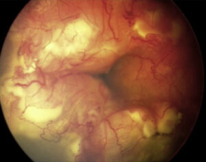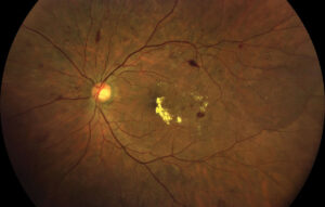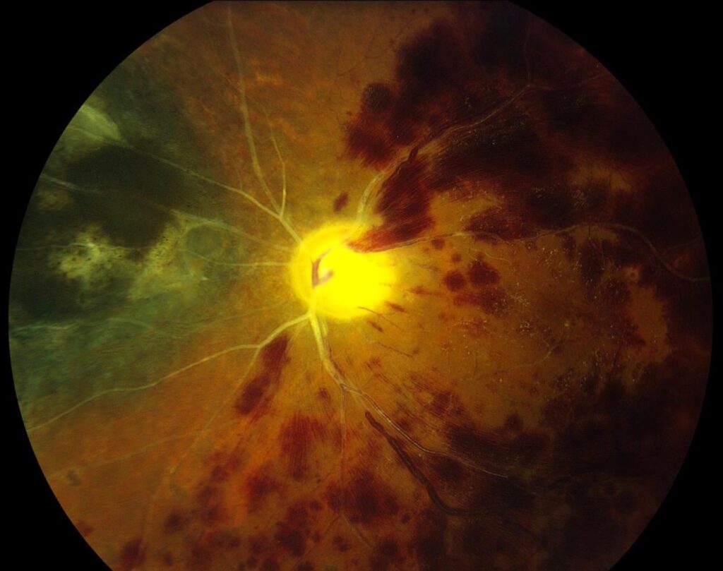
Radiation retinopathy treatment aims to preserve central vision after plaque brachytherapy, proton beam or external beam radiation near the eye.
The disease is an occlusive microangiopathy that usually appears months to years after exposure, so management follows a stepwise plan based on dose, timing and macular involvement. For clinical signs and imaging, see our radiation retinopathy review.
Why Radiation Retinopathy Treatment Is Difficult
Radiation injures capillary endothelial cells, and perfusion loss progresses slowly, so as a result ischemic retina keeps releasing VEGF long after the tumor has responded. Current therapy suppresses edema and neovascularization, although it cannot revive closed capillaries. Patients should hear this early, because expectations shape adherence.
Who Is at Risk
Risk rises with total dose, dose to the fovea and optic disc, and tumor proximity to the macula. In addition, diabetes, hypertension and concurrent chemotherapy appear to increase susceptibility. Plaque therapy for posterior uveal melanoma accounts for many eyes in published series.
Fundus Explorer Pro
Photograph the retinal findings described here with the phone already in your pocket — 22 D optics and built-in illumination in one handheld unit.
From Choroida — the team behind this siteBaseline Assessment
First, document acuity, fundus photographs and OCT before or soon after radiation, and next, record the tumor margin relative to the fovea and disc. Fluorescein angiography then maps nonperfusion once retinopathy appears. OCT angiography may show capillary dropout before visible hemorrhages, while its prognostic value is still uncertain.
Prophylaxis in Radiation Retinopathy Treatment
Several groups tested prevention in eyes with high predicted macular dose. For example, a randomized trial found that periocular triamcinolone at plaque placement, 4 months and 8 months reduced macular edema and moderate vision loss at 18 months.
In addition, bevacizumab every 4 months reduced macular edema in a large series compared with historical controls. Sector laser to the irradiated retina also showed benefit in pilot data. However, much of this evidence is nonrandomized, so protocols differ between centers.
Anti-VEGF in Radiation Retinopathy Treatment
Intravitreal anti-VEGF is the usual first-line therapy for radiation maculopathy, and bevacizumab, ranibizumab and aflibercept all appear in published series, mostly retrospective. Injections reduce macular thickness and can stabilize acuity in many eyes, but edema often recurs, and long-term series describe injections continuing over years. The approach mirrors diabetic macular disease (see DME treatment compared).
Injection Intervals and Follow-Up
Most clinicians start with monthly loading and then extend intervals by OCT response. Treat-and-extend suits this disease because recurrence is common and unpredictable, and clinicians also review the tumor at each visit, since oncology surveillance continues in parallel.
Steroids in Radiation Retinopathy Treatment
Steroids target the inflammatory component that anti-VEGF may miss, and intravitreal triamcinolone reduced radiation macular edema in an early series, although the effect was often temporary. Some retina specialists use the dexamethasone implant off-label for longer action in refractory eyes. However, cataract and pressure rise limit repeated dosing, and therefore steroids fit pseudophakic eyes or patients who cannot attend frequent visits.
Laser in Radiation Retinopathy Treatment
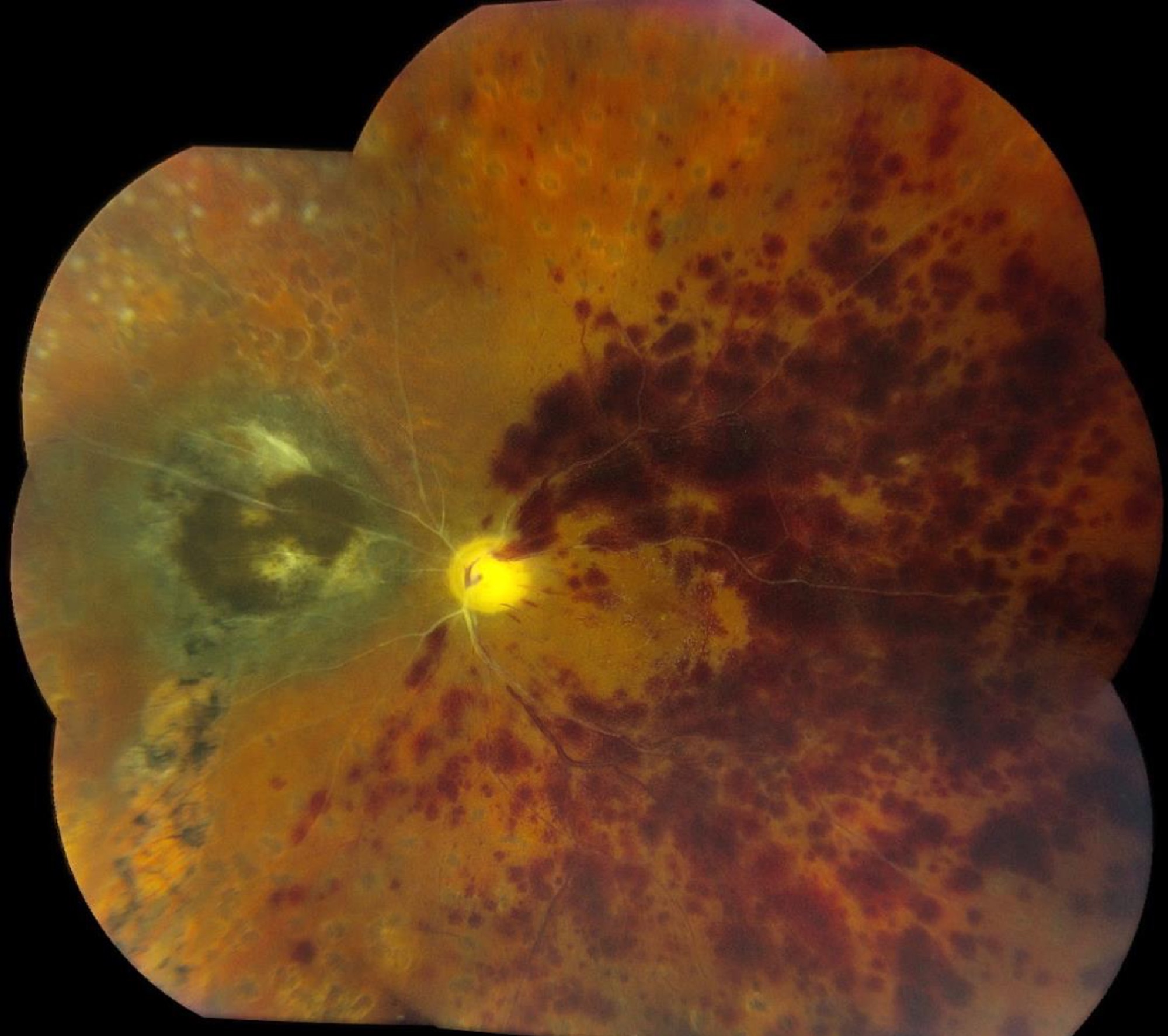
Scatter photocoagulation treats neovascularization of the disc, retina or iris. It reduces the ischemic drive and lowers the risk of vitreous hemorrhage and neovascular glaucoma. Focal laser for macular edema now has a smaller role because anti-VEGF works faster.
Combination Therapy
Many refractory eyes receive anti-VEGF and a steroid together or in sequence, and then sector laser may reduce the injection burden. Evidence comes from small series, so tailor the plan to response and side effects.
Proliferative Disease and Complications
Nonclearing vitreous hemorrhage may need vitrectomy with endolaser, while neovascular glaucoma requires anti-VEGF, panretinal laser and pressure control, sometimes with a drainage device. Radiation optic neuropathy can coexist and carries a guarded prognosis.
What Radiation Retinopathy Treatment Cannot Do
No current option reperfuses closed capillaries, and as a result, eyes with established foveal ischemia rarely regain vision despite a dry OCT. Check the foveal avascular zone before predicting improvement.
Counseling Patients
Explain that the disease is chronic and may need injections over a long period, and also stress that tumor control comes first and that stable vision is a realistic goal. Patients should report new floaters, distortion or eye pain promptly.
Pitfalls to Avoid
- Do not mistake radiation exudation for tumor recurrence without imaging review
- Avoid observing center-involving edema while vision declines
- Never repeat steroids without checking pressure and lens status
- Do not promise visual gain when angiography shows foveal ischemia
- Remember that neovascular glaucoma can develop with little retinal hemorrhage


Document what you see
Two smartphone imaging tools built for everyday clinic use — one for the slit lamp, one for the fundus.
From Choroida — the team behind this siteReferences
- Brown GC, Shields JA, Sanborn G, et al. Radiation retinopathy. Ophthalmology. 1982;89:1494-1501.
- Horgan N, Shields CL, Mashayekhi A, et al. Periocular triamcinolone for prevention of macular edema after plaque radiotherapy of uveal melanoma: a randomized controlled trial. Ophthalmology. 2009;116:1383-1390.
- Shah SU, Shields CL, Bianciotto CG, et al. Intravitreal bevacizumab at 4-month intervals for prevention of macular edema after plaque radiotherapy of uveal melanoma. Ophthalmology. 2014;121:269-275.
- Finger PT, Chin KJ, Semenova EA. Intravitreal anti-VEGF therapy for macular radiation retinopathy: a 10-year study. Eur J Ophthalmol. 2016;26:60-66.
- Shields CL, Demirci H, Dai V, et al. Intravitreal triamcinolone acetonide for radiation maculopathy after plaque radiotherapy for choroidal melanoma. Retina. 2005;25:868-874.
- Materin MA, Bianciotto CG, Wu C, Shields CL. Sector laser photocoagulation for the prevention of macular edema after plaque radiotherapy of uveal melanoma: a pilot study. Retina. 2012;32:1601-1607.

Writes and reviews the clinical articles on Educate. Every post is checked against peer-reviewed sources before it goes live.