DME prognosis depends on how much damage the macula already carries when treatment starts.
Anti-VEGF therapy dries most maculae, yet final vision varies widely between patients. Therefore, the clinician needs predictors that go beyond central thickness, because OCT layers, baseline acuity, chronicity and systemic control all shape the outlook.
What Determines DME Prognosis
Visual outcome reflects two separate processes. First, fluid distorts the retina, and this distortion is largely reversible, while second, neuronal and photoreceptor loss accumulates and does not recover with drying. As a result, two eyes with identical thickness can end with very different acuity.
Baseline Visual Acuity
Baseline acuity predicts both the gain and the final level, and in DRCR Retina Network trials, eyes starting with worse vision gained more letters on average. However, they usually finished with lower absolute acuity than eyes that started better, so a large gain does not promise normal reading vision.
Fundus Explorer Pro
Photograph the retinal findings described here with the phone already in your pocket — 22 D optics and built-in illumination in one handheld unit.
From Choroida — the team behind this siteCentral Thickness and DME Prognosis
Central subfield thickness guides retreatment, but it correlates only modestly with acuity. For example, a dry macula can still see poorly when inner layers are disorganized, so avoid quoting thickness alone as the measure of success.
DRIL as a Predictor of DME Prognosis
Disorganization of the retinal inner layers (DRIL) means the ganglion cell, inner plexiform, inner nuclear and outer plexiform boundaries become indistinguishable. Sun and colleagues measured its horizontal extent within the central 1 mm, and greater DRIL extent was associated with worse acuity in center-involved DME, while early resolution of DRIL was also associated with later visual improvement.
Ellipsoid Zone Integrity
The ellipsoid zone reflects photoreceptor inner segment health, and disruption of this band under the fovea is associated with poorer acuity in DME. Maheshwary and colleagues found that greater percent disruption tracked with worse vision. Furthermore, an intact external limiting membrane suggests that photoreceptors may recover after fluid resolves, so assess these outer bands on a high-quality foveal scan before counseling.
Chronicity in DME Prognosis
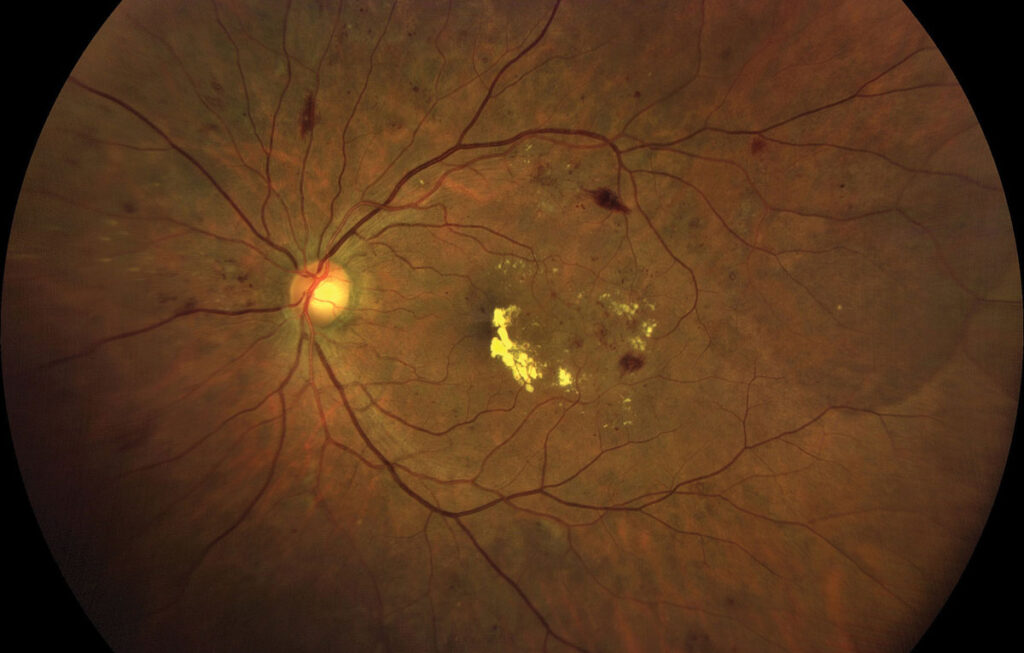
Edema that persists for months or years damages the retina structurally, and long-standing cysts can leave inner retinal thinning and outer retinal atrophy after drying. Similarly, chronic leakage deposits subfoveal hard exudate that may organize into fibrosis, so delayed referral often limits the ceiling for recovery.
Early Response and DME Prognosis
The first few injections offer useful prognostic information, and in a post hoc analysis of Protocol I, eyes with limited early gain generally showed limited long-term gain. However, some limited early responders still improved with continued therapy, so early response refines expectations but should not end treatment by itself.
Choice of Anti-VEGF Agent
Protocol T compared aflibercept, bevacizumab and ranibizumab, and at one year, aflibercept gave greater mean gain when baseline acuity was 20/50 or worse. With better baseline vision, mean differences between agents were small, and drug choice therefore matters most in eyes with worse starting vision.
Persistent Edema
Some eyes keep fluid despite regular monthly anti-VEGF injections, but persistent edema does not always mean poor vision, and many of these eyes still gain letters. Nevertheless, switching to a corticosteroid may help selected eyes (see steroids in resistant DME).
Systemic Factors in DME Prognosis
Glycemic control, blood pressure, lipids and kidney function influence the retinal course. Poor attendance also commonly undermines outcomes in routine practice, so adherence counseling forms part of the prognosis discussion.
Macular Ischemia
FAZ enlargement and parafoveal capillary loss limit visual recovery, and OCT angiography demonstrates these areas without dye injection. When ischemia involves the fovea, injections may dry the macula without restoring acuity, so document perfusion when vision lags behind anatomy (see diabetic macular ischemia).
Framing Expectations in DME Prognosis
Patients usually ask whether vision will return to normal, so first, explain that treatment aims to stabilize vision, with improvement possible in many eyes. Next, link the expected outcome to the patient’s own OCT findings. For example, intact outer bands and limited DRIL support a hopeful message, while extensive DRIL or ellipsoid zone loss calls for a cautious forecast.
Pitfalls to Avoid
- Do not promise normal vision based on a dry OCT alone
- Do not ignore DRIL and ellipsoid zone loss when acuity lags
- Avoid stopping treatment after a weak early response without review
- Never overlook ischemia, cataract or epiretinal membrane as competing causes
- Remember that systemic control and visit adherence shape the outcome


Document what you see
Two smartphone imaging tools built for everyday clinic use — one for the slit lamp, one for the fundus.
From Choroida — the team behind this siteReferences
- Sun JK, Lin MM, Lammer J, et al. Disorganization of the retinal inner layers as a predictor of visual acuity in eyes with center-involved diabetic macular edema. JAMA Ophthalmol. 2014;132:1309-1316.
- Wells JA, Glassman AR, Ayala AR, et al. Aflibercept, bevacizumab, or ranibizumab for diabetic macular edema. N Engl J Med. 2015;372:1193-1203.
- Gonzalez VH, Campbell J, Holekamp NM, et al. Early and long-term responses to anti-vascular endothelial growth factor therapy in diabetic macular edema: analysis of Protocol I data. Am J Ophthalmol. 2016;172:72-79.
- Maheshwary AS, Oster SF, Yuson RM, et al. The association between percent disruption of the photoreceptor inner segment-outer segment junction and visual acuity in diabetic macular edema. Am J Ophthalmol. 2010;150:63-67.
- Bressler SB, Qin H, Beck RW, et al. Factors associated with changes in visual acuity and central subfield thickness at 1 year after treatment for diabetic macular edema with ranibizumab. Arch Ophthalmol. 2012;130:1153-1161.

Writes and reviews the clinical articles on Educate. Every post is checked against peer-reviewed sources before it goes live.